Rat neural cultures showing strong neuron specific induction of c-Fos following depolarization using our mouse monoclonal antibody.
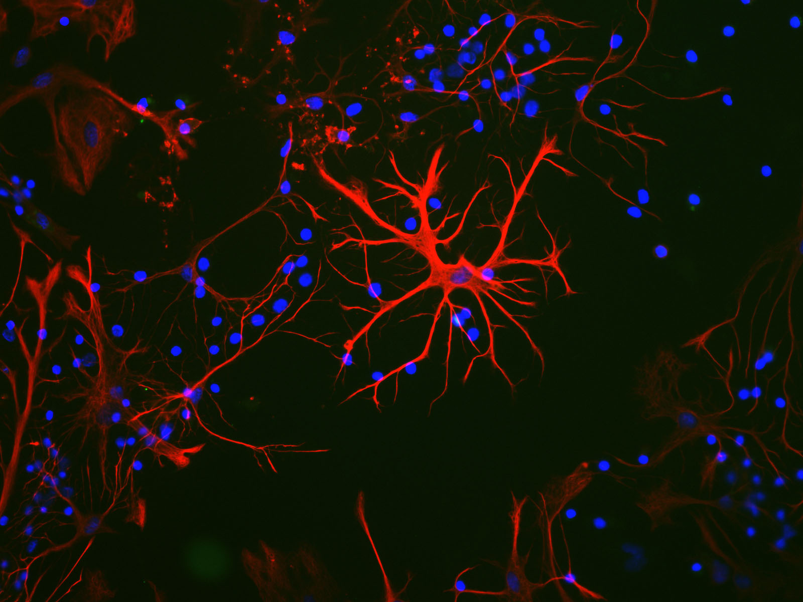
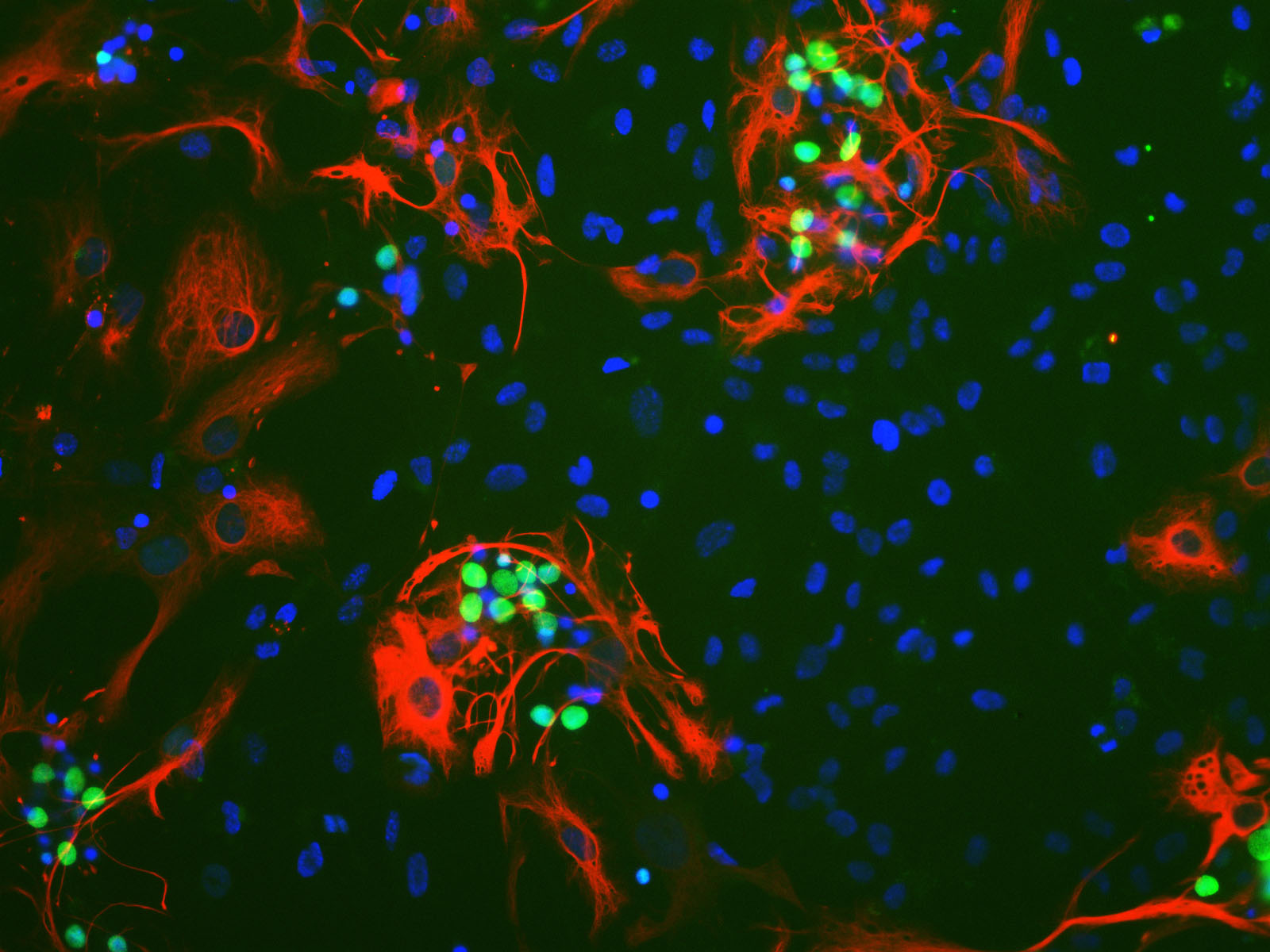
Rat brain neural cultures (left) and the same cells stimulated with membrane deplorization buffer for 5 hours (right). This is a salt solution containing 170mM Potassium which depolarizes and stimulates gene expression in neuronal cells but has no effect on glia. Cultures were stained with our monoclonal antibody to c-Fos, MCA-2H2 in green and rabbit anti-GFAP, RPCA-GFAP in red. Nuclear DNA is revealed
with DAPI in blue. The c-Fos expression is strongly induced only in stimulated neuronal cells and localizes in the nucleus, while unstimulated neurons and glia show no c-Fos staining. The c-Fos antibody was used at a dilution of
1:1,000 from a 1mg/mL solution. The GFAP antibody was used at a
dilution of 1:1,000. Cultures were processed using our standard fixation
and staining procedure (described here).
To order this antibody go to our order form (here) or check out from the antibody page here.
Picture taken with a Zeiss 40X objective and documented with a SPOT camera. Mouse click on each image to get an enlarged view.