Double Label Immunofluorescence with Antibodies to Vimentin and GFAP
Rat glial cells in mixed cerebral cortex cultures stained with chicken antibody to Vimentin (green) and counterstained with rabbit antibody to GFAP (red). Hoechst dye reveals nuclear DNA in blue. Link here to the EnCor Biotechnology Home Page
.
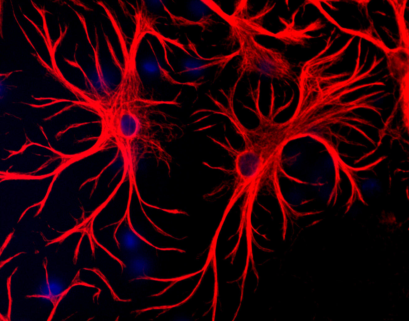
Rat cerebral
cortex cultures stained with chicken
antibody to vimentin (green) and rabbit
antibody to GFAP (red). Note flattened fibroblastic cells are mostly
green (i.e. vimentin positive, GFAP negative), while clearly astrocytic
cell, such as the one in top center, express both vimentin and GFAP and
therefore appear golden or orange. Certain other cells express predominantly
GFAP and therefore appear red. Specimen processed using our standard protocols
(described here).
To order either of these antibodies go to our order form (here).
Picture taken with a Zeiss 40X objective and documented with a Diagnostic Instruments Digital
SPOT camera.