Neurons in tissue culture stained with out polyclonal rabbit c-Fos antibody showing strong c-Fos induction in depolarized neurons.
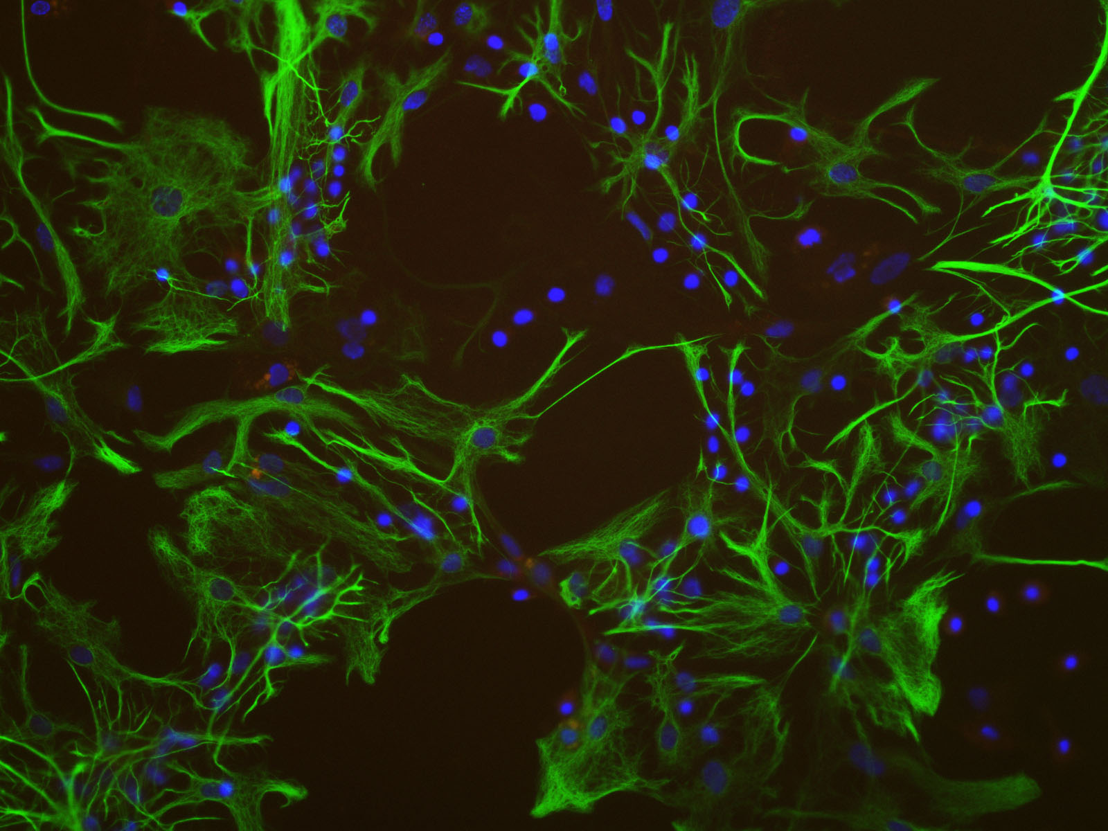
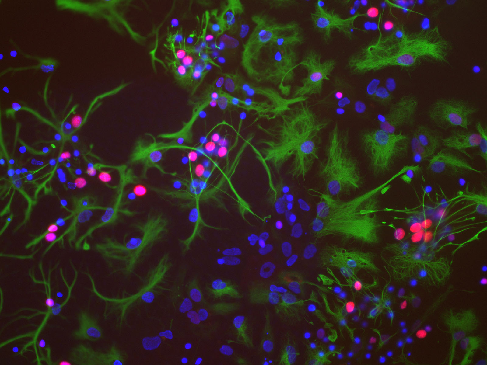
Left: Rat brain mixed neural cultures were stained with our polyclonal antibody to c-Fos, RPCA-c-Fos-AP in red and Monoclonal antibody to GFAP, MCA-5C10 in green. Nuclear DNA is revealed with DAPI in blue. Right: A similar culture stimulated with membrane depolarization buffer for 5 hours and stained with same antibodies as in left panel. This buffer is rich in Potassium and depolarizes and stimulates neurons but not non-neuronal cells. Strong c-Fos expression is seen in nuclei of the neurons is this culture but not in the nuclei of glia. The c-Fos antibody was used at a dilution of 1:1,000 from purified antibody at 1mg/mL. The GFAP antibody was used at a dilution of 1:1,000. Cultures were processed using our standard fixation
and staining procedure (described here).
To order this antibody go to our order form (here) or go to the c-fos antibody page and use our online store here.
Picture taken with a Zeiss 40X objective and documented with a SPOT camera. Mouse click on each image to get an enlarged view.