Data on Neuromics Hippocampal Neurons
We obtained Neuromics E18 Primary Rat Hippocampal Cells, which arrived unfrozen in a ~2ml screw top tube. We were interested in using these cells to stain them with our antibodies to get some nice images, and we figured that would be good for both EnCor and Neuromics. However we can also say that we did this work independently, and if the cells had not performed as advertized you would not be reading this page. Following more or less the Neuromics protocol we coated 35mm sterile tissue culture dishes with 0.1mg/ml polylysine for one hour, washed them with distilled water, allowed them to dry and sterilized them by leaving them under a UV light in a sterile hood. The Neuromics protocol first treats the hippocampal cells for 30 minutes with papain in B27 culture media- the B27 is supplied in the kit, but the papain is not. This step dissociates the cells, and is followed by mild centrifugation to allow the removal of the papain. We then plated the cells out in the B27/Neurobasal medium supplied in the kit, and incubated them in our CO2 incubator. After three days we saw numerous beautiful hippocampal neurons and by seven days there were long neuronal processes anastomosizing through the cultures. We fixed and stained the cultures using some of our battery of monoclonal and polyclonal antibodies using our standard tissue culture cell staining protocol, details of which are here. We were able to test these cells with various of our neuronal and stem cell markers and found them to be positive for vimentin, UCHL1, α-synuclein, α-internexin, Tau, Doublecortin, NF-L, NF-M, MAP2, Fox3/NeuN, GAP43 and α-II spectrin. Some of the results are shown below.

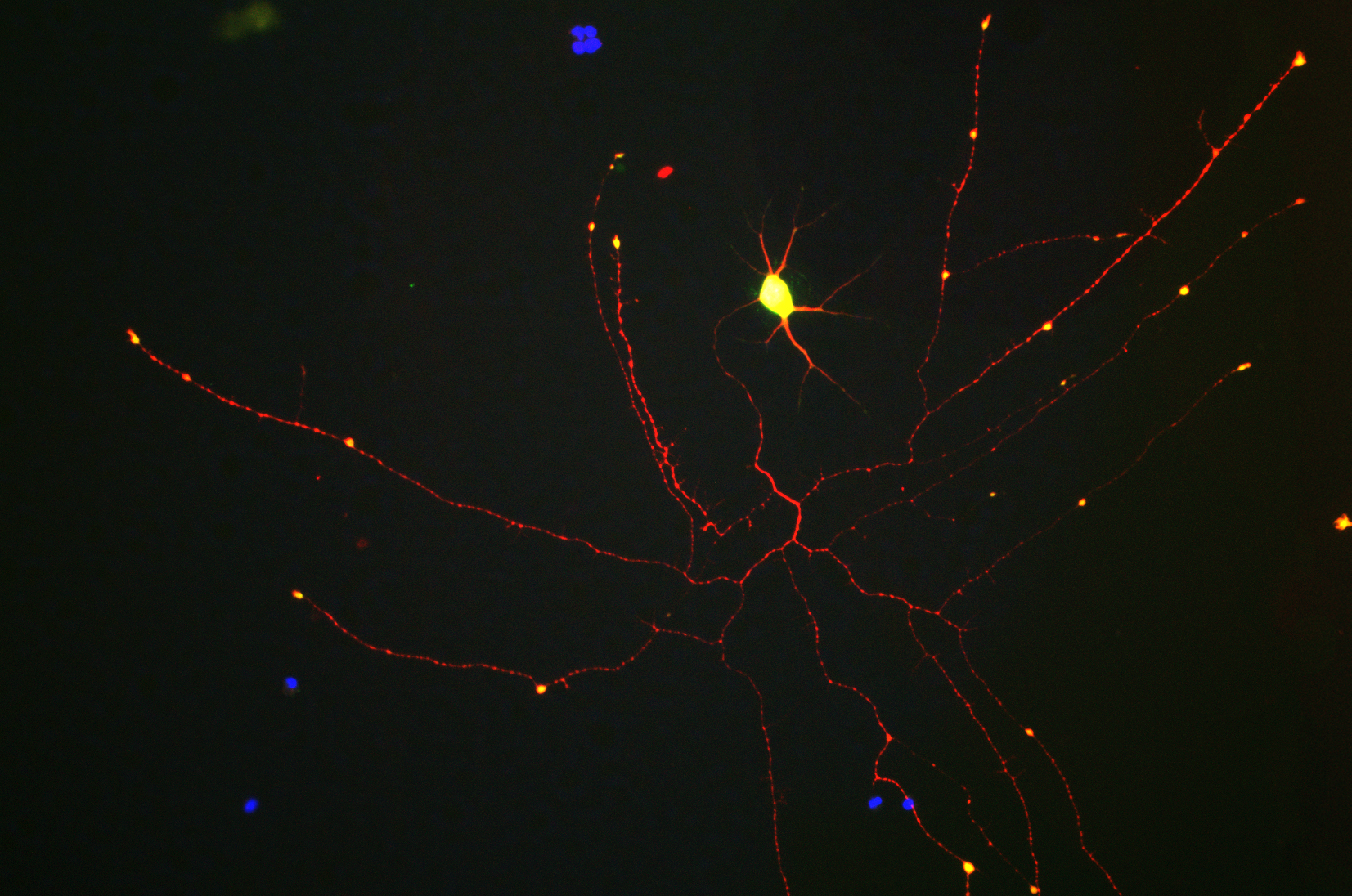
Legend: Neuromics E18 hippocampal neurons grown for 7 days in tissue culture and stained with CPCA-Tau, EnCor's chicken polyclonal antibody to microtubule associated protein tau (red) and Doublecortin antibody MCA-3E1 (green). The two proteins overlap in the proximal dendrites, but doublecortin is more abundant in the growth cones and periphery. As a result, the periphery appears green while the more proximal regions of the cells are yellow. The single longer process of this cell, presumably an axon, has a low doublecortin content and so appears red. Blue staining is the nuclear DNA.
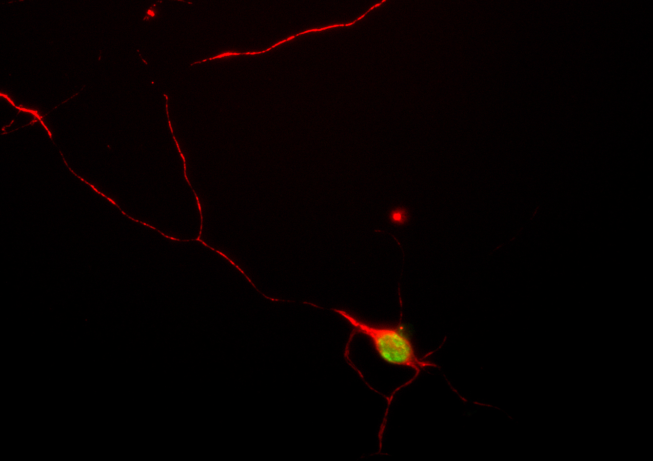
Legend: Another view of Neuromics E18 hippocampal neurons grown for seven days and stained in the red channel with CPCA-Tau, our polyclonal antibody to microtubule associated protein tau, an abundant protein of neuronal perikarya and axons. The cells were also stained in green with MCA-BH7, our monoclonal antibody to UCHL1, an abundant cytoplasmic protein of neurons which is concentrated in the perikarya. Since the perikarya contain both UCHL1 and Tau, the red and green signals superimpose, giving a yellow color, while the process, which contain relatively much more tau, appear red. Note that the growth cones, that active migrating tips of the neuronal processes, are yellowish indicating considerable accumulation of UCHL1 there also. Blue stain is DAPI and reveals cell nuclei of some non neuronal cells in these cultures.

Legend: Another view of Neuromics E18 hippocampal neurons grown for seven days and stained in the red channel with CPCA-Int, our polyclonal antibody to the neurofilament subunit α-internexin which forms short filaments in these cells at this stage. The cells were also stained in green with MCA-2E9, our monoclonal antibody to microtubule associated protein tau, an abundant cytoplasmic protein of neurons which is found in the perikarya, dendrites and axon. Blue stain is DAPI and reveals cell nuclei of neurons and some non neuronal cells in these cultures.
Legend: Another view of Neuromics E18 hippocampal neurons grown for four days and stained in the red channel with CPCA-NF-M, our polyclonal antibody to the neurofilament subunit NF-M which forms short filaments in these cells at this stage. The cells were also stained in green with MCA-39C7, our pan specific monoclonal antibody recognizing nuclear pore complexes.
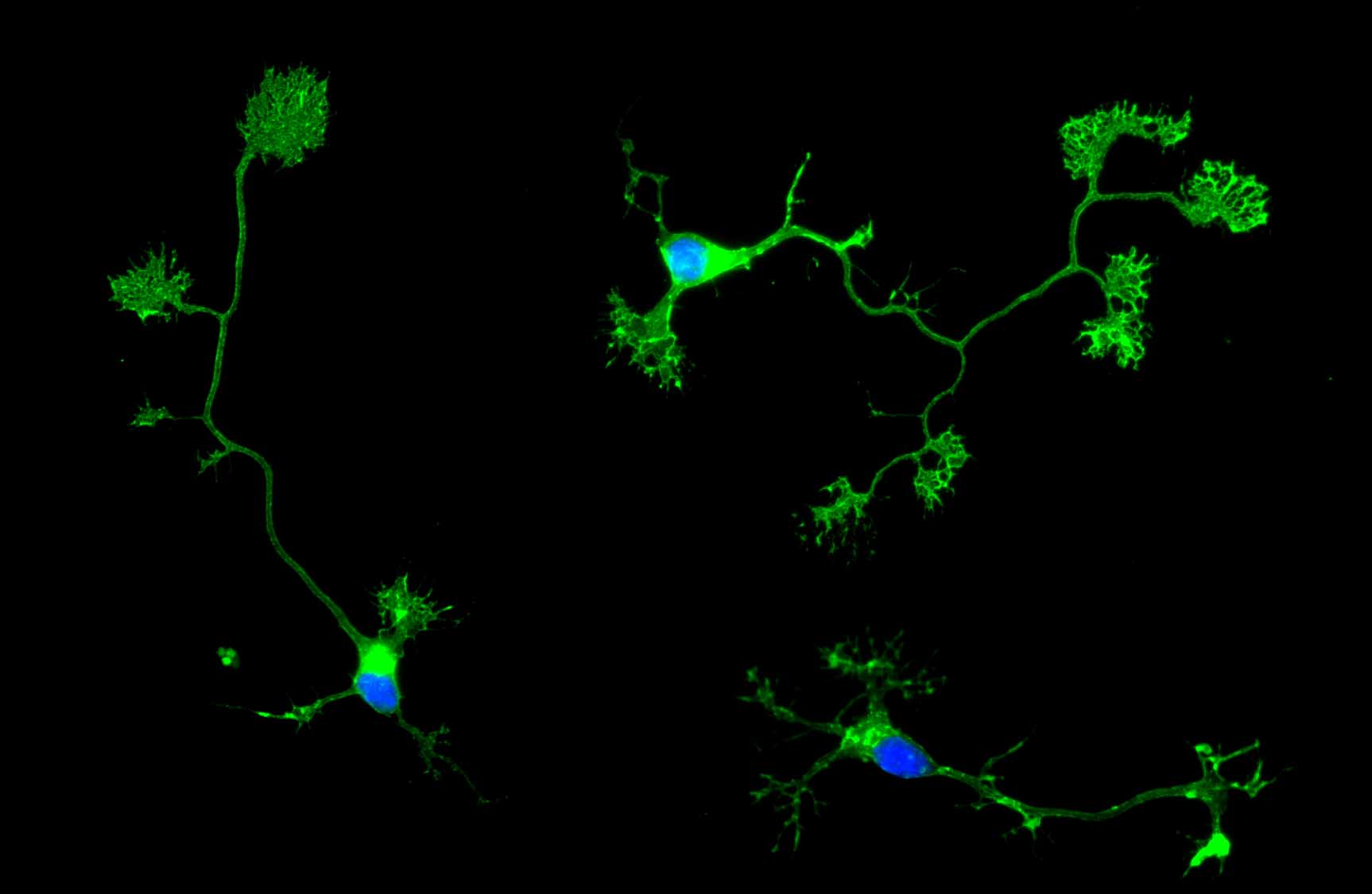
Legend: Neuromics E18 hippocampal neurons grown for four days and stained in the green channel with MCA-1E3, our monoclonal antibody to Growth Associated Protein 43 (GAP43) an abundant protein of neuronal membranes especially concentrated in axonal membranes and growth cones. GAP43 is heavily palmitoylated in the Golgi, so this antibody can be used to localize the neuronal Golgi complex. Blue stain is DAPI and reveals cell nuclei of these neuronal cells.
Use of Images or Text: The contents of this page are available for modification and reuse under the terms of the Creative Commons Attribution/Share-Alike License 3.0 and the GNU Free Documentation License, unversioned with no invariant sections, front-cover texts, or back-cover texts. These licences permit modification and reuse, even commercially, as long as authorship credit and a link to this page is given.