Redirecting to new URL....
Catalogue# CPCA-mCherry: Chicken Polyclonal Antibody to mCherryThe Immunogen: mCherry is derived from proteins originally isolated from Cnidarians (jelly fish, sea anemones and corals), and is used as a fluorescent tracer in trasfection and transgenic experiments. The prototype for these fluorescent proteins is Green Fluorescent Protein (GFP), which is a ~27 kDa protein isolated originally from the jellyfish Aequoria victoria. GFP was the basis of the 2008 Nobel Prize in Chemistry, awarded to Osamu Shimomura, Martin Chalfie and Roger Tsien, specifically for "for the discovery and development of the green fluorescent protein, GFP". GFP was shown to fluoresce on contact with molecular oxygen, requiring no other cofactors, and so can be expressed in fluorescent form in essentially any prokaryotic or eukaryotic cell. The mCherry protein is derived from DsRed, a red fluorescent protein from so-called disc corals of the genus Discosoma. DsRed is similar in size and properties to GFP, but, obviously, produces a red rather than a green fluorochrome. The original DsRed was engineered extensively in the Tsien lab to prevent it from forming tetramers and dimers and to modify and improve the spectral properties (1-3). Several further cycles of mutation, directed modification and evolutionary selection produced mCherry, which has an excitation maximum at 587 nm and and emission maximum at 610 nm (4). We expressed the mCherry protein sequence shown in reference 4 in bacteria, purified out the mCherry and raised a rabbit polyclonal antibody. This was affinity purified and was found to stain a band of the expected size in Hek293 cells transfected with a vector designed to express mCherry which was obtained from Clontech. We also market a rabbit polyclonal antibody and a mouse monoclonal antibody to mCherry, specifically RPCA-mCherry and MCA-1C51.
We are OEM suppliers of this antibody- in other words we manufactured it, characterized it and generated the data presented on this page. This antibody is available from several other vendors, but we can supply it more cheaply and we can provide you with more detailed information on the properties of the antibody.
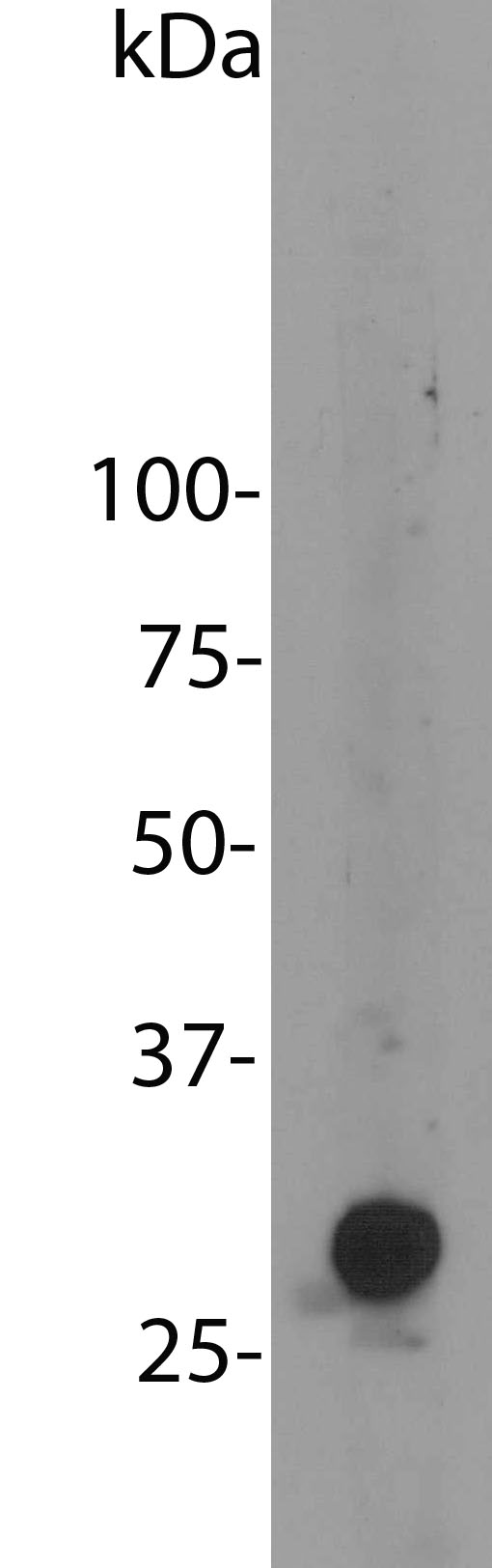
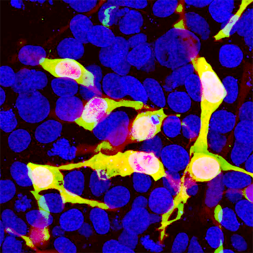
Figures: Left: Blot of HEK293 cells transfected with pFin-EF1-mCherry vector, courtesy of the Semple-Rowland lab at the University of Florida. There is a strong clean band at about 30 kDa. HEK293 cells which were not transfected with this vector show no protein bands. Right: HEK293 cells transfected in the same way and viewed in the confocal microscope. Most HEK293 cells are not transfected so only the nucleus of these cells can be visualized with a blue DNA stain. Cells which are transfected with mCherry are bright red. Staining with CPCA-mCherry is shown in Green. Green antibody staining is only seen cells which express Cherry, as expected, and the superimposition of the green and red results in an orange signal. Interestingly stronger Cherry staining is seen in the nucleus, possibly due to degradation of some Cherry molecules to release the low molecular weight Cherry fluorochrome.
Antibody Characteristics: Antibody was raised in chicken against recombinant full length His tagged mCherry purified from E. coli. This antibody is an IgY prep at a total protein concentration of 30 mg/mL. The preparation contains 10mM sodium azide as a preservative. Store at 4°C or -20°C. Avoid repeat freezing and thawing.
Suggestions for use: Try at dilutions of 1:1,000 and higher for immunofluorescence. For western blots try at 1:2,000. The mCherry protein runs at about 30kDa on SDS-PAGE gels.
Omim link: No OMIM link, this is an engineered version of an invertebrate protein not found in humans.
References:
1. Baird GS, Zacharias DA, Tsien RY. Biochemistry, mutagenesis, and oligomerization of DsRed, a red fluorescent protein from coral. Proc Natl Acad Sci U S A. 97:11984-9 (2000).
2. Gross LA, Baird GS, Hoffman RC, Baldridge KK, Tsien RY. The structure of the chromophore within DsRed, a red fluorescent protein from coral. Proc Natl Acad Sci U S A. 97:11990-5 (2000).
3. Heikal AA, Hess ST, Baird GS, Tsien RY, Webb WW. Molecular spectroscopy and dynamics of intrinsically fluorescent proteins: coral red (dsRed) and yellow (Citrine). Proc Natl Acad Sci U S A. 97:11996-2001 (2000).
4. Shaner NC, Campbell RE, Steinbach PA, Giepmans BN, Palmer AE, Tsien RY. Improved monomeric red, orange and yellow fluorescent proteins derived from Discosoma sp. red fluorescent protein. Nature Biotechnology 22:1567-1572 (2004).
Limitations: This product is for research use only and is not approved for use in humans or in clinical diagnosis.
Availability and Price: Available for shipping now, $200 US per aliquot of 100 μL of IgY antibody, enough for hundreds of experiments. For order form press here.
Use of Images or Text: The contents of this page are available for modification and reuse under the terms of the Creative Commons Attribution/Share-Alike License 3.0 and the GNU Free Documentation License, unversioned with no invariant sections, front-cover texts, or back-cover texts. These licences permit modification and reuse, even commercially, as long as authorship credit and a link to this page is given.