Redirecting to new URL....
Catalogue CPCA-MAP2: Chicken Polyclonal Antibody to Microtubule Associated Protein 2 (MAP2)
The Immunogen: Microtubules are 25nm diameter protein rods found in most kinds of eukarytic cells. They are polymerized from a dimeric subunit made of one a subunit and one b tubulin subunit. Microtubules are associated with a family of proteins called microtubule associated proteins (MAPs), which includes the protein t (tau) and a group of proteins referred to as MAP1, MAP2, MAP3, MAP4 and MAP5. MAP2 is made up of two ~280kDa apparent molecular weight bands referred to as MAP2a and MAP2b. A third lower molecular weight form, usually called MAP2c, corresponds to a pair of protein bands running at ~70kDa on SDS-PAGE gels. All these MAP2 forms are derived from a single gene by alternate transcription, and all share a C-terminal sequence which includes either three or four microtubule binding peptide sequences, which are very similar to those found in the related microtubule binding protein t (tau). MAP2 isoforms are expressed only in neuronal cells and specifically in the perikarya and dendrites of these cells. Antibodies to MAP2 are therefore excellent markers on neuronal cells, their perikarya and neuronal dendrites. In contrast t (tau) is found predominantly in neuronal axons. The HGNC name for this protein is MAP2.
We are OEM suppliers of this antibody- in other words we manufactured it, characterized it and generated the data presented on this page. This antibody is available from several other vendors, but we can supply it more cheaply and we can provide you with more detailed information on the properties of the antibody.
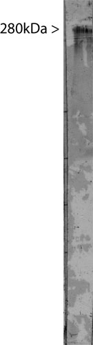
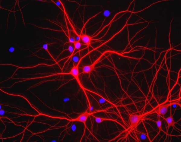
Left: Western blot of extract of bovine brain stained with CPCA-MAP2, revealing a band at 280kDa. Right: View of mixed neuron/glial cultures stained with CPCA-MAP2 (red). The perikarya and dendrites of neurons are strongly and specifically stained with the MAP2 antibody, while the axons of the neurons and the processes of all other cell types in these cultures (astrocytes, oligodendrocytes, microglia, endothelia and fibroblasts) are all negative. Cell nuclei are visualized with DAPI DNA stain.
Antibody Characteristics: This antibody was generated in chicken by
standard procedures and immunoglobulin was extracted from egg yolk. The resulting
polyclonal antibody belongs to the IgY subclass. This is the chicken homologue
of mammalian IgG and can be used in the same general way, with the caveat that
this type of antibody does not bind either Protein A or Protein G. Suitable
second antibody reagents can be obtained from many vendors including Molecular
Probes and Sigma-Aldrich. Store at 4°C or -20°C. Avoid repeat freezing and thawing.
Suggestions for use: The IgY solution is at a concentration of ~19mg/ml and has an extremely high titre against MAP2. It can be used at dilutions of 1:10,000 - 1:20,000 in immunofluorescence experiments.
Omim link: press here.
References:
1. Goetz AK, Scheffler B, Chen HX, Wang S, Suslov O, Xiang H, Brüstle O, Roper SN, Steindler DA. Temporally restricted substrate interactions direct fate and specification of neural precursors derived from embryonic stem cells. Proc Natl Acad Sci U S A. 103:11063-11068 (2006).
2. Walton NM, Snyder GE, Park D, Kobeissy F, Scheffler B, Steindler DA. Gliotypic neural stem cells transiently adopt tumorigenic properties during normal differentiation. Stem Cells 27:280-289 (2009).
Price and Availability: - We currently supply 50 microliter aliquots for $250. Material is in stock and ready for immediate shipping.
Limitations: This product is for research use only and is not approved for use in humans or in clinical diagnosis.
Use of Images or Text: The contents of this page are available for modification and reuse under the terms of the Creative Commons Attribution/Share-Alike License 3.0 and the GNU Free Documentation License, unversioned with no invariant sections, front-cover texts, or back-cover texts. These licences permit modification and reuse, even commercially, as long as authorship credit and a link to this page is given.
©EnCor Biotechnology Inc. .