Catalogue# MCA-BH7: Mouse Monoclonal Antibody to Ubiquitin C-Terminal Hydrolase 1
The Immunogen: Ubiquitin C-terminal hydrolase 1 (UCHL1) has several other names, such as ubiquitin carboxyl esterase L1, ubiquitin thiolesterase, neuron-specific protein PGP9.5 and Park5. It was originally identified as a major component of the neuronal cytoplasm from 2-dimensional gel analysis of brain tissues, and was given the name PGP9.5 (1). The protein is extremely abundant, and was estimated to be present at a concentration of 200-500 micrograms/g wet weight, representing a major protein component of neuronal cytoplasm (1). This has been claimed to represent 1-2% of total brain protein (2). It was later found that a ubiquitin C-terminal hydrolase enzyme activity was associated with the PGP9.5 protein, resulting in the renaming of PGP9.5 to UCHL1. This is the first of a family of ubiquitin C-terminal hydrolases which have been characterized, many of which also have rigid cell type specific expression patterns. The ubiquitin C-terminal hydrolases cleave ubiquitin from other molecules. This activity is important to generate mono-ubiquitin from the several genes which encode polyubiquitin chains or ubiquitin fused to other proteins. The activity is also important to remove ubiquitin from partially degraded proteins, allowing the ubiquitin monomer to be recycled. Regulation of the ubiquitin pathway is very important and many disease states are associated with defects in this pathway. For example the Park5 gene causes one form of human Parkinson's disease, and proves to be a point mutations in the UCHL1 gene producing a I93M form of the UCHL1 protein which has reduced ubiquitin hydrolase activity (3). Interestingly a common allelic variant of UCHL1, the S18Y polymorphism is actually protective against Parkinson's disease. Recent studies suggest that UCHL1 also has a ubiqutinyl ligase activity, being able to couple ubiquitin monomers by linking the C-terminus of one with lysine 63 of the other (3). Since UCHL1 is heavily expressed in neurons, antibodies to UCHL1 can be used to identify neurons in histological sections and in tissue culture. The great abundance of this protein in neurons means that it is released from neurons in large amounts following injury or degeneration, so the detection of UCHL1 in CSF and other bodily fluids can be used as a biomarker of neuronal injury or degeneration. The HGNC name for this protein is UCHL1.
We are OEM suppliers of this antibody- in other words we manufactured it, characterized it and generated the data presented on this page. This antibody is available from several other vendors, but we can supply it more cheaply and we can provide you with more detailed information on the properties of the antibody.
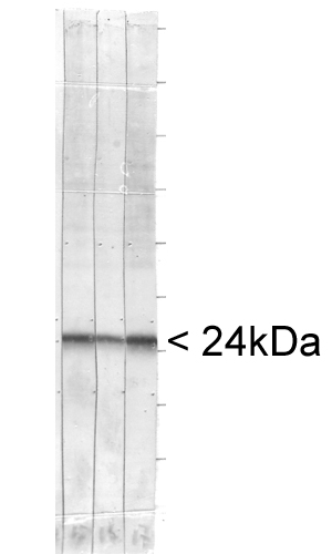
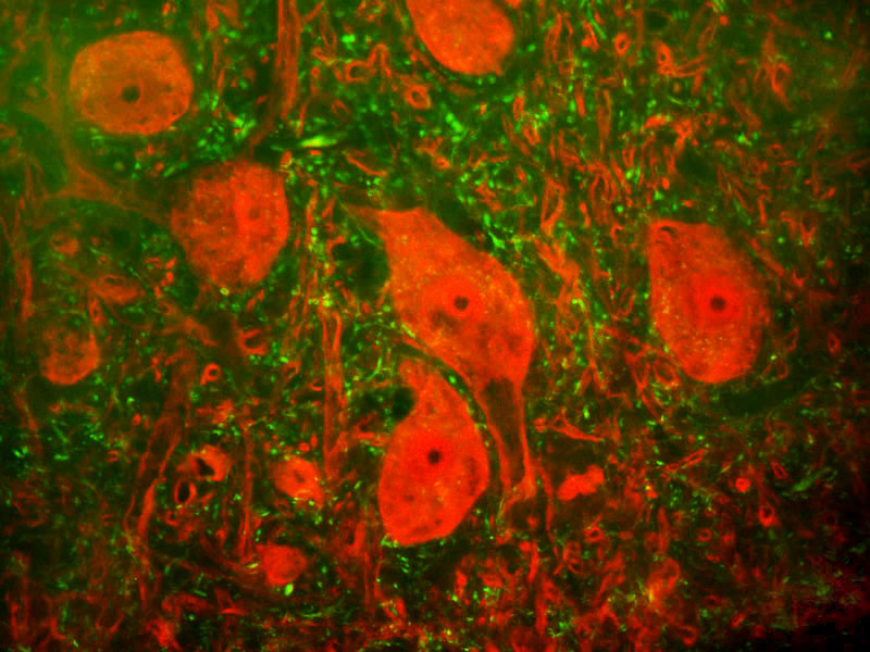
Figures: Left: Blots of whole cell homogenate of bovine brain stained with MCA-BH7 (right most lane) and two other monoclonal antibodies reactive with UCHL1 (left and central lane). All three antibodies show a single clean band running at about 24kDa. Right: shows a section of rat spinal cord stained with UCHL1 clone MCA-BH7 (red) and RPCA-NF-H,
EnCor's rabbit antibody to neurofilament NF-H (green). The large cells are a-motorneurons and UCHL1 fills the cytoplasm of their perikarya and dendrites. The neurofilament NF-H antibody binds primarily to phosphorylated axonal forms of NF-H, and so stains axons coursing between the large motor neurons.
Antibody Characteristics: Antibody was raised in mouse against recombinant full length human UCHL1 purified from E. coli. This antibody is in the form of 100 microliters of ascites fluid which are typically over 1mg/ml specific antibody. The antibody is an IgG1 subclass. The preparation contains 10mM sodium azide as a preservative. Store at 4°C or -20°C. Avoid repeat freezing and thawing.
Suggestions for use: Try at dilutions of 1:2,000 for immunofluorescence. For Western blots try at 1:20,000. A suitable control tissue is rat spinal cord, brain, SHSY-5Y or HEK293 cell extract. The UCHL1 protein runs at about 24kDa on SDS-PAGE gels, and is a prominent component of brain, spinal cord and especially cortical extracts.
OMIM Link: press here
References:
1. Doran JF, Jackson P, Kynoch PA, Thompson RJ. Isolation of PGP 9.5, a new human neurone-specific protein detected by high-resolution two-dimensional electrophoresis. J Neurochem. 40:1542-7 (1983).
2. Wilkinson KD, Lee KM, Deshpande S, Duerksen-Hughes P, Boss JM, Pohl J. The neuron-specific protein PGP 9.5 is a ubiquitin carboxyl-terminal hydrolase. Science. 1989 246:670-3 (1989).
3. Liu Y, Fallon L, Lashuel HA, Liu Z, Lansbury PT Jr. The UCH-L1 gene encodes two opposing enzymatic activities that affect alpha-synuclein degradation and Parkinson's disease susceptibility. Cell 111:209-18 (2002).
Limitations: This product is for research use only and is not approved for use in humans or in clinical diagnosis.
Availability and Price: Available for shipping now, $200 US per aliquot of 100 microliters of ascites fluid, enough for hundreds of experiments. For order form press here
Use of Images or Text: The contents of this page are available for modification and reuse under the terms of the Creative Commons Attribution/Share-Alike License 3.0 and the GNU Free Documentation License, unversioned with no invariant sections, front-cover texts, or back-cover texts. These licences permit modification and reuse, even commercially, as long as authorship credit and a link to this page is given.