Data on Neuromics hN2 Neuronal Stem Cells
The hN2 cells are human neuronal lineage cells which originate from ArunA Biomedical, and were derived from the human embryonic cell line WA09. We obtained hN2 Human Neurons Discovery kit from Neuromics, which arrived frozen and on dry ice. We were interested in using these cells to stain them with our antibodies to get some nice images, and we figured that would be good for both EnCor and Neuromics. However we can also say that we did this work independently, and if the cells had not performed as well as advertized you would not be reading this page. Following more or less the Neuromics protocol we coated 35mm sterile tissue culture dishes with Matrigel diluted with 10 volumes of Dulbecco's modified Eagle medium (DME) culture media under aseptic conditions. We made up the hippocampal culture media from 97mls of AB2 media and 2mls of ANS supplement, both included in the kit. To this were added 100 microliters of leukemia inhibitory factor (LIF) at 10 micrograms/ml, 1ml of 200mM L-Glutamine and 1ml of penicillin/streptomycin, which were not included in the kit. After 2 days we saw many clumps of cells from which long processes were emanating. We fixed and stained the cultures using some of our battery of monoclonal and polyclonal antibodies using our standard tissue culture cell staining protocol, details of which are here. We were able to test these cells with various of our neuronal and stem cell markers and found them to be positive for vimentin, UCHL1, α-synuclein, NF-L, MAP2 and
-II spectrin. They were apparently negative for NeuN/Fox3. Some of the results are shown below.
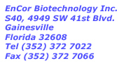
Legend: Neuromics hN2 cells stained with our chicken polyclonal antibody to vimentin CPCA-Vim, in red. Islands of Hn2 cells form after 4 days in culture forming beautiful flower like structures. Vimentin is a well established marker of early differentiating neuronal lineage cells, though it is found in many other kinds of cells. Taken with a 10X objective lens. Blue staining is the nuclear DNA.
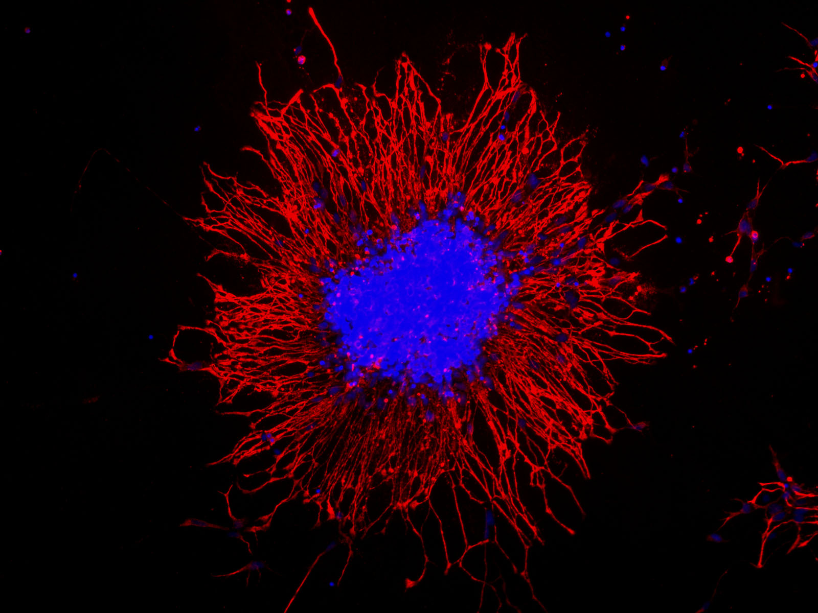
Legend: Neuromics hN2 cells grown in culture for 4 days and stained with our chicken polyclonal to MAP2, CPCA-MAP2, a marker of neuronal dendrites and perikarya. Differentiating cells show strong cytoplasmic staining for MAP2. Blue stain is DAPI and reveals cell nuclei in these cultures.
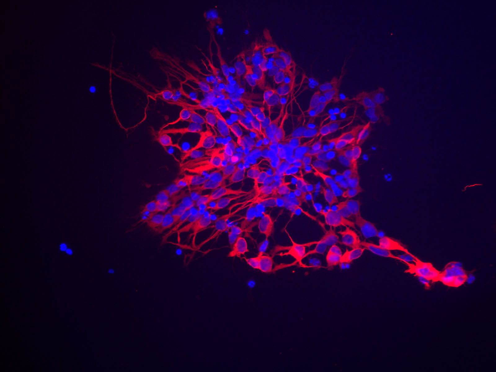
Legend: Neuromics hN2 cells grown in culture for 4 days and stained with our chicken polyclonal to neurofilament light or low molecular weight chain NF-L, CPCA-NF-L, a marker of neurons. Many of the differentiating cells show strong cytoplasmic and clearly fibrillar staining for NF-L. Blue stain is DAPI and reveals cell nuclei in these cultures.
Use of Images or Text: The contents of this page are available for modification and reuse under the terms of the Creative Commons Attribution/Share-Alike License 3.0 and the GNU Free Documentation License, unversioned with no invariant sections, front-cover texts, or back-cover texts. These licences permit modification and reuse, even commercially, as long as authorship credit and a link to this page is given.