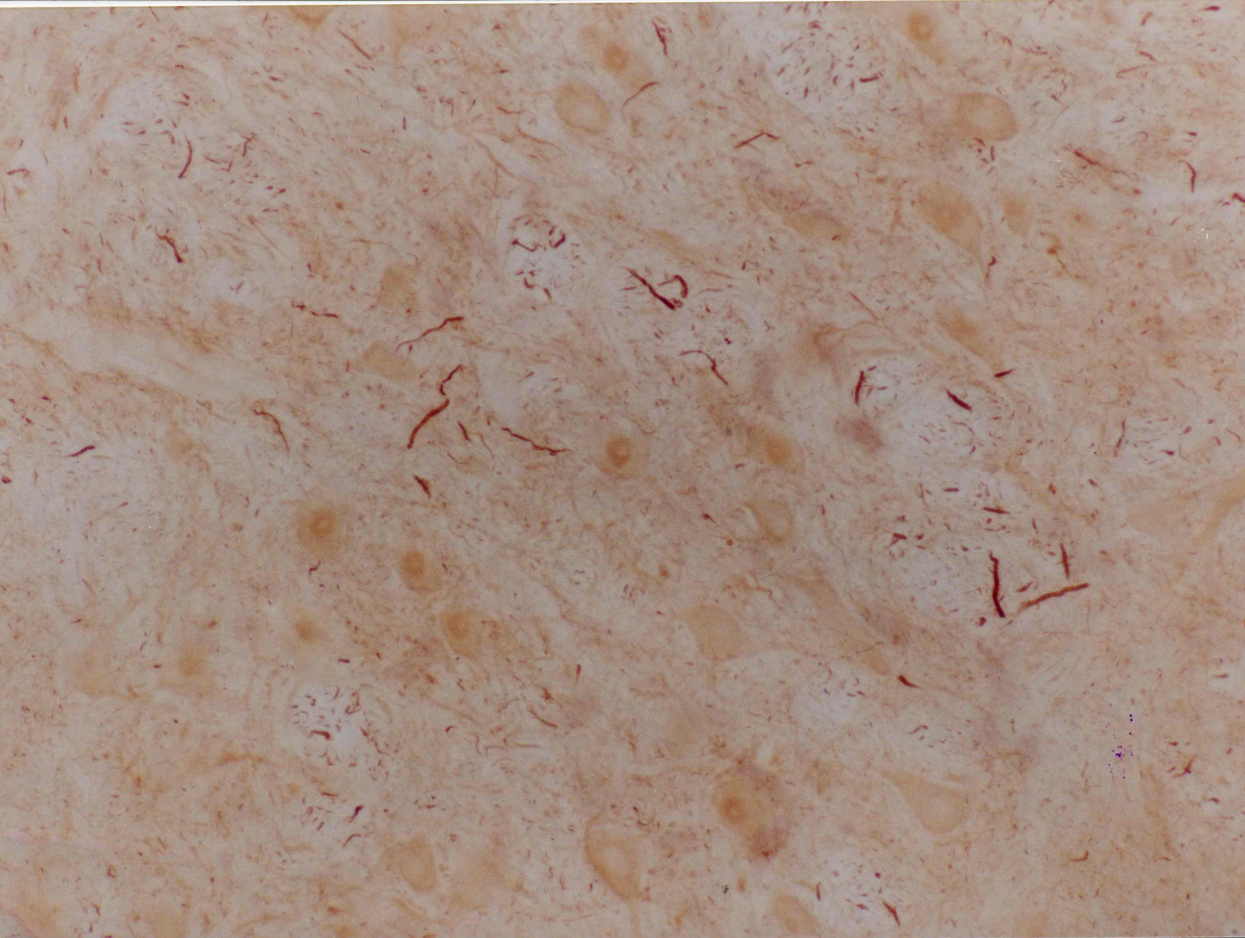
Sections of adult rat brain stem showing contral lateral facial nucleus to that shown in previous figure, also 3 days after lesion of the contralateral facial nerve. Stained using ABC-DAB technique with 2E3 monoclonal antibody to a-internexin. Note strong staining only of fibres passing through the nucleus. There is very little cytoplasmic staining of the neuronal cell bodies. Link here back to the EnCor Biotechnology Home Page.

Sections of adult rat brain stem showing contralateral facial nucleus 3 days after lesion of the facial nerve and stained using ABC-DAB technique with 2E3 monoclonal antibody to a-internexin. Note lack of strong cytoplasmic staining in neuronal cell bodies. To order this antibody go to our order form (here). Picture taken with a Zeiss 40X objective and documented with a SPOT camera.