October 2023 News
We now have new data on two rabbit polyclonal antibodies to microtubule associated protein 2, RPCA-MAP2A/B and a new antibody RPCA-MAP2D. The MAP2 gene produces several different sized proteins by alternate transcription. MAP2C and MAP2D are lower molecular weight isotypes which are expressed early in development and are around 70kDa molecular size when run on SDS-PAGE. MAP2A and MAP2B isotypes are expressed later in development and on SDS-PAGE run at about 220kDa, due to the inclusion of large sequence inserts which produce the MAP2 “projection domains”. Projection domains can be seen in the electron microscope as fine protrusions from the side of microtubules in neuronal perikary and dendrites. Since MAP2C and MAP2D sequences are included in the MAP2A and MAP2B the new RPCA-MAP2D antibody will recognize all forms of MAP2, while RPCA-MAP2A/B will recognize only the mature forms of the protein. Both antibodies work well on western blots, IF, ICC and IHC.
We are currently gearing up the the Society for Neuroscience Meetimg in Washington DC, 11th to 15th November. Come and visit us at Booth 139 which is easy to remember as it is 30, 31, 32. We will be giving out the usual pens, posters, flashlights, postcards and antibody samples, and also making available more of our Santiago Ramon-Y-Cajal and Camillo Golgi laser cut images so see you there!
September 2023 News
We now have new data on two rabbit polyclonal antibodies to microtubule associated protein 2, RPCA-MAP2A/B and a new antibody RPCA-MAP2D. The MAP2 gene produces several different sized proteins by alternate transcription. MAP2C and MAP2D are lower molecular weight isotypes which are expressed early in development and are around 70kDa molecular size when run on SDS-PAGE. MAP2A and MAP2B isotypes are expressed later in development and on SDS-PAGE run at about 220kDa, due to the inclusion of large sequence inserts which produce the MAP2 “projection domains”. Projection domains can be seen in the electron microscope as fine protrusions from the side of microtubules in neuronal perikary and dendrites. Since MAP2C and MAP2D sequences are included in the MAP2A and MAP2B the new RPCA-MAP2D antibody will recognize all forms of MAP2, while RPCA-MAP2A/B will recognize only the mature forms of the protein. Both antibodies work well on western blots, IF, ICC and IHC.
We are currently gearing up the the Society for Neuroscience Meetimg in Washington DC, 11th to 15th November. Come and visit us at Booth 139 which is easy to remember as it is 30, 31, 32. We will be giving out the usual pens, posters, flashlights, postcards and antibody samples, and also making available more of our Ramon-Y-Cajal and Golgi laser cut images so see you there!
August 2023 News
We release yet more novel antibodies. We made a novel rabbit polyclonal and a mouse mpnoclonal to the enzyme catalase, RPCA-Catalase and MCA-6H14, both good markers of peroxisomes. As we always do, we document how they work on western blots for IF, ICC and IHC and show the relevant data.
June 2023 News
We have now finished testing all our rabbit polyclonal antibodies for immunohistochemistry on formalin fixed and paraffin embedded (FFPE) rodent and rodent tissues. So we found that many of them work very well, as shown in the examples below. So now we can recommend (or not) all of our antibodies on FFPE human and rodent specimens, describe the protocols we used and show convincing example images.
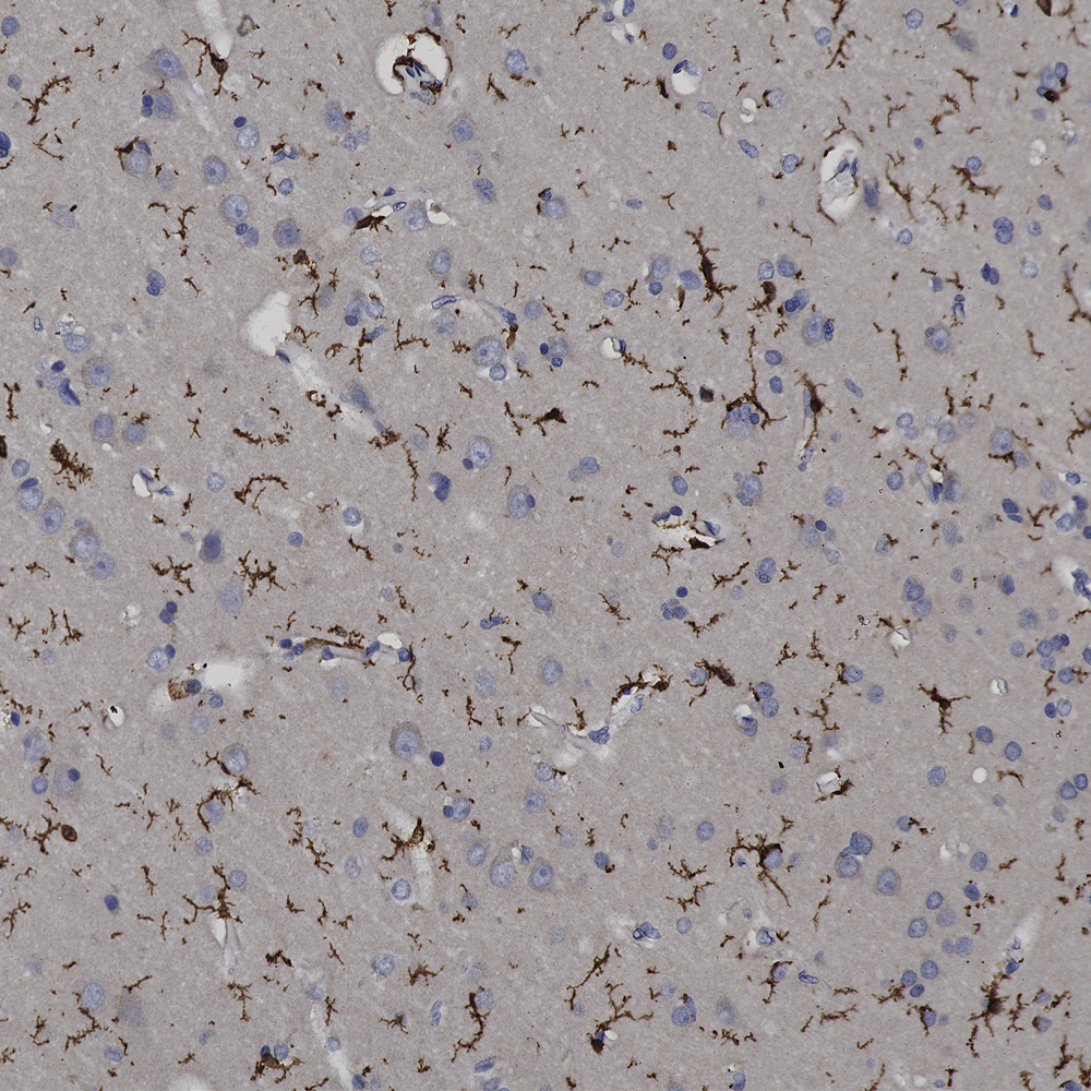
Chromogenic immunostaining of a NBF fixed paraffin embedded human brain cortex section with rabbit pAb to IBA1, RPCA-IBA1, dilution 1:2,000, detected with DAB (brown) using the Vector Labs ImmPRESS method and reagents with citra buffer retrieval. Hematoxylin (blue) was used as the counterstain. The RPCA-IBA1 antibody specifically labels the cytoplasm of microglial cells. This antibody performs well in testing with 4% PFA and standard NBF fixed mouse, rat and human tissue.
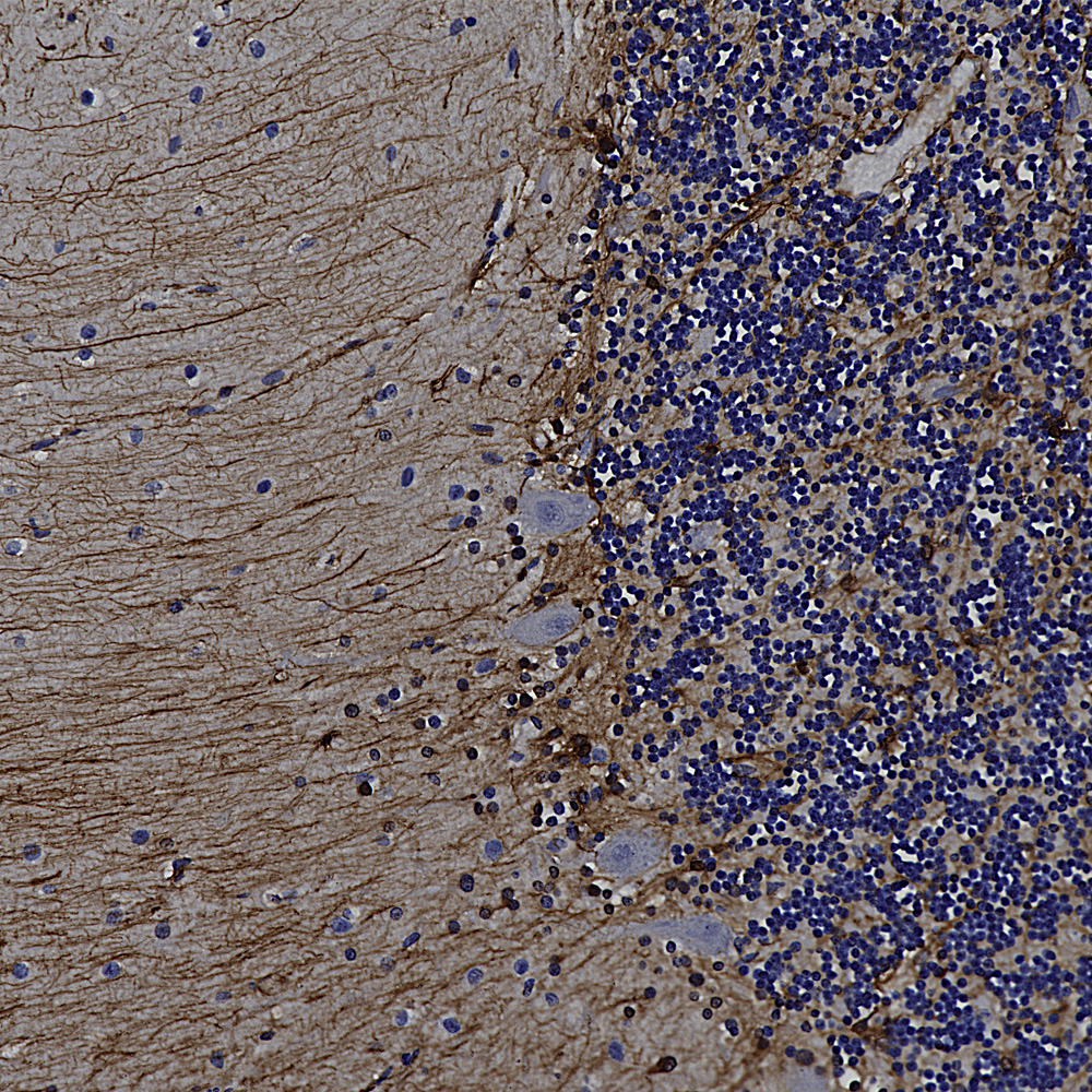
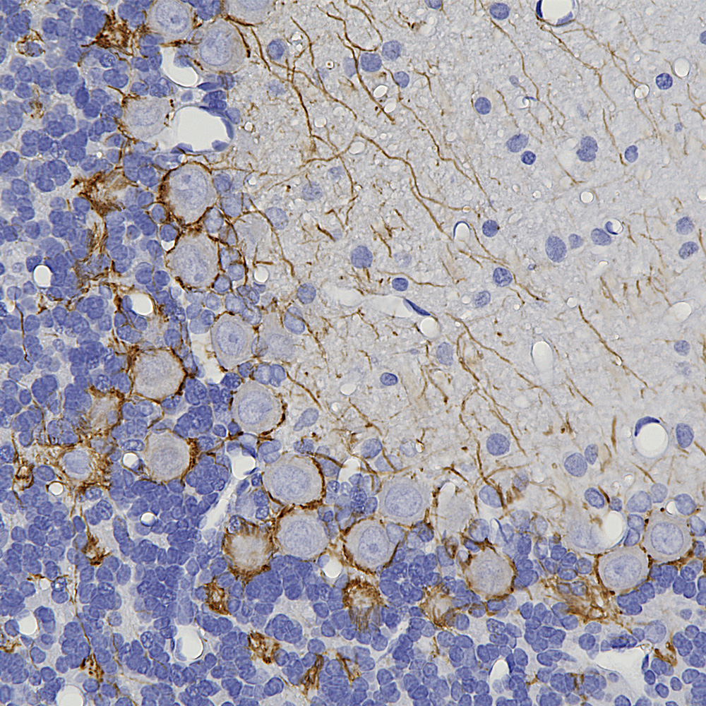
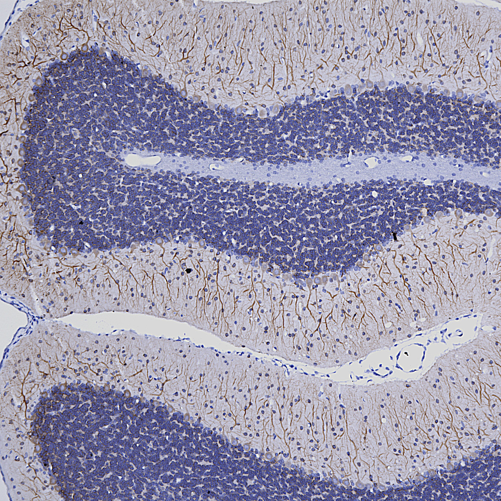
Chromogenic immunostaining of a formalin fixed paraffin embedded human cerebellum section with rabbit pAb to GFAP, RPCA-GFAP, dilution 1:5,000, detected with DAB (brown) using the Vector Labs ImmPRESS method and reagents with citra buffer retrieval. Hematoxylin (blue) was used as the counterstain. The RPCA-GFAP antibody labels the processes of astrocytes within both the granular and molecular layers and Bergmann glia in the molecular layer. This antibody performs well in testing with both 4% PFA and standard NBF fixed rat, mouse, and human tissues.
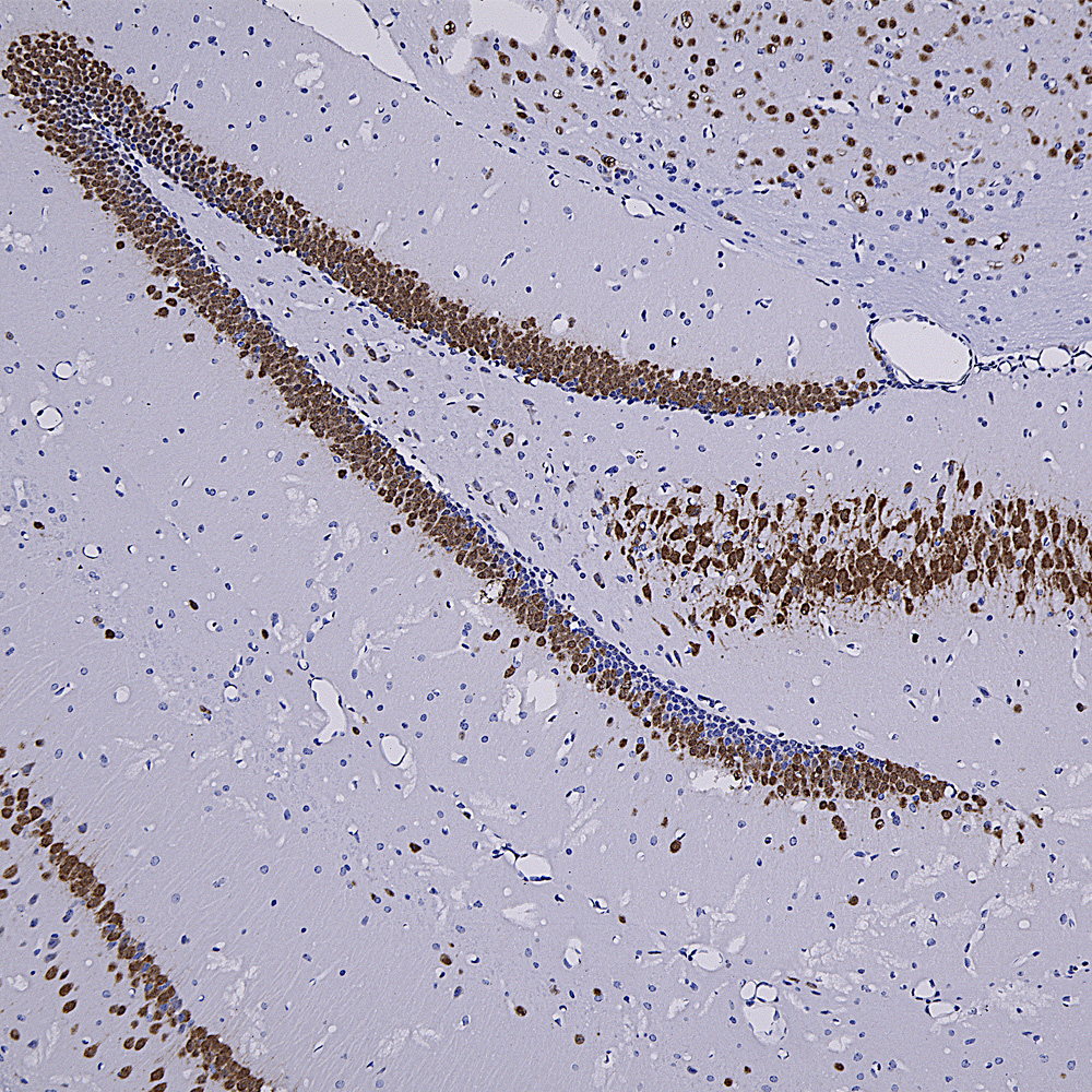
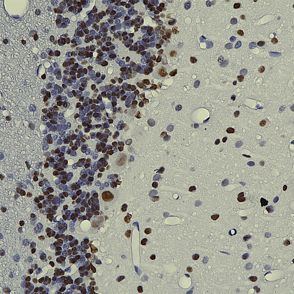
Chromogenic immunostaining of a formalin fixed paraffin embedded mouse hippocampus section with rabbit pAb to Fox3/NeuN, RPCA-FOX3, dilution 1:4,000, detected with DAB (brown) using the Vector Labs ImmPRESS method and reagents with citra buffer retrieval. The RPCA-FOX3 antibody selectively labels the nuclei and distal perikaryal of most neuronal cell populations. This antibody performs well in testing with 4% PFA and NBF fixed mouse, human, and rat tissues. Mouse select image for larger view.
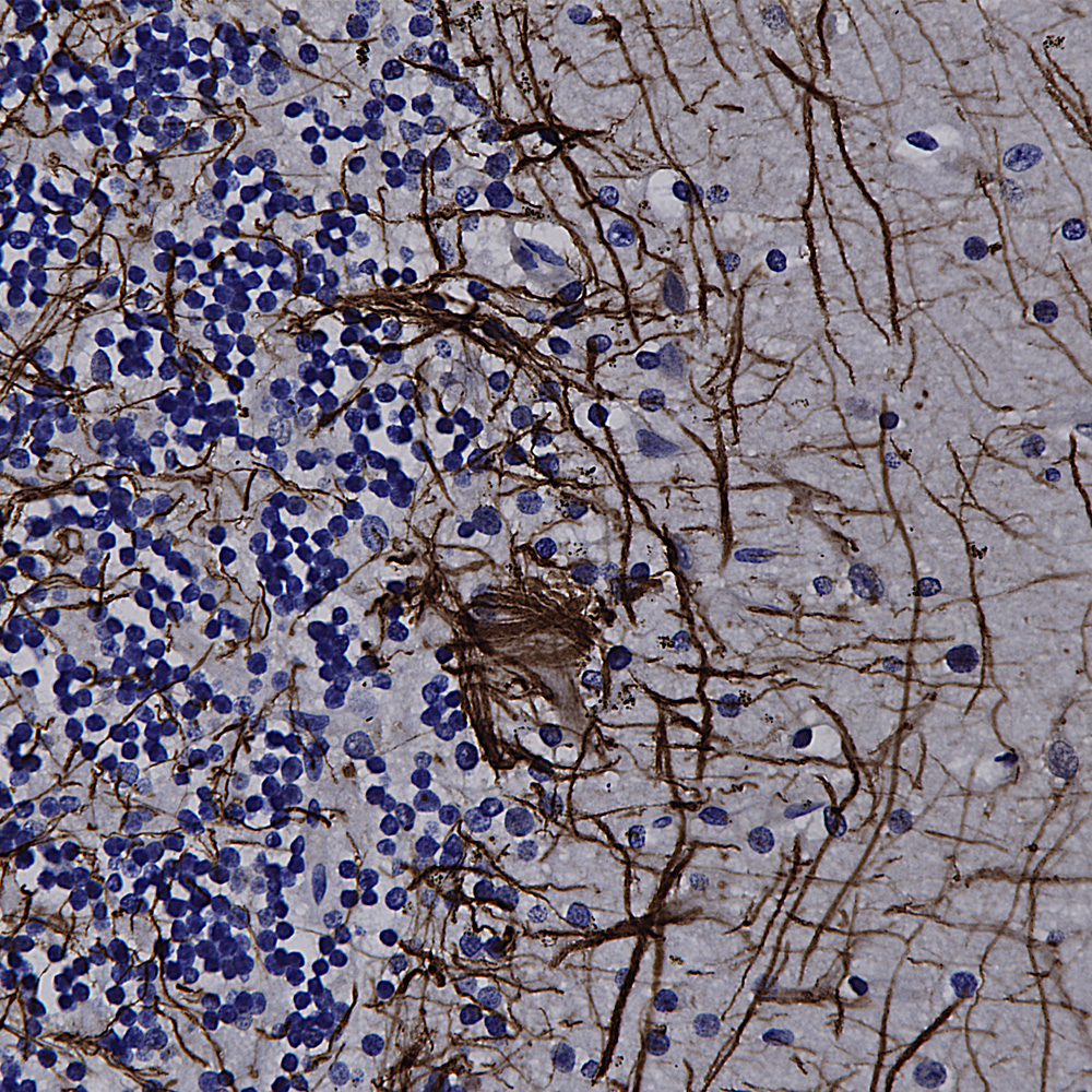
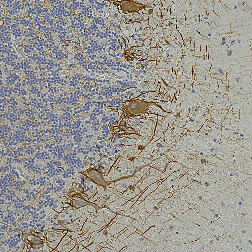
Chromogenic immunostaining of a formalin fixed paraffin embedded human cerebellum section with rabbit pAb to NF-L, RPCA-NF-L-ct, dilution 1:5,000, detected with DAB (brown) using the Vector Labs ImmPRESS method and reagents with citra buffer retrieval. Hematoxylin (blue) was used as the counterstain. RPCA-NF-L-ct strongly labels the axons and dendrites of Purkinje cells and the projections of neuronal cells within the granular layer. This antibody performs well in testing with both 4% PFA and standard NBF fixed rat, mouse, and human tissues. Mouse select image for larger view.
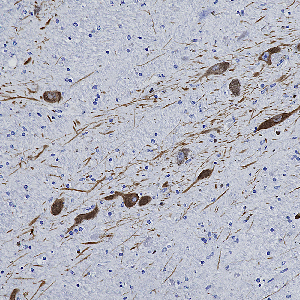
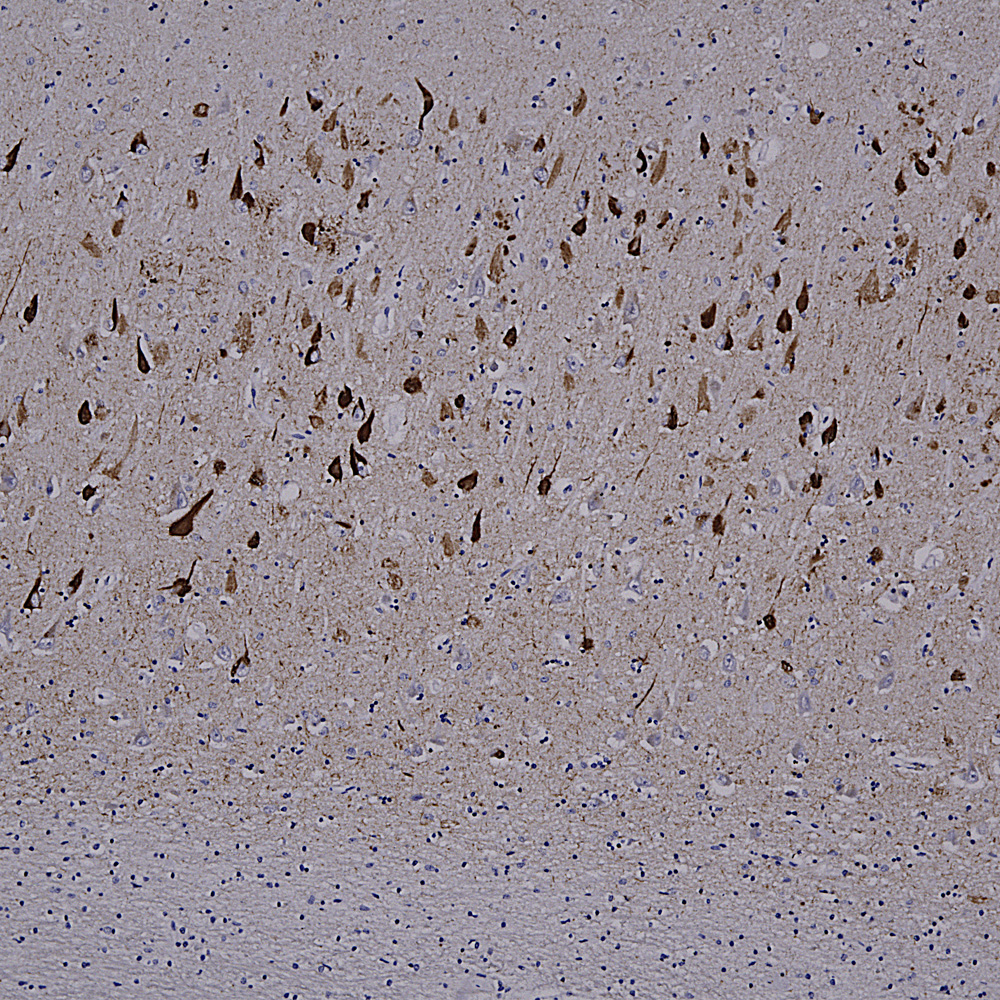
Chromogenic immunostaining of a NBF fixed paraffin embedded human midbrain section with rabbit pAb to tyrosine hydroxylase, RPCA-TH, dilution 1:10,000, detected with DAB (brown) using the Vector Labs ImmPRESS method and reagents with citra buffer retrieval. Hematoxylin (blue) was used as the counterstain. In this image, RPCA-TH antibody labels dopaminergic neurons and their axons traversing the striatum. This antibody performs well in testing with 4% PFA and standard NBF fixed mouse, rat and human tissue. Mouse select image for larger view.
April 2023 News
Our recent work on the Uman NF-L antibodies published in pre-print form in BioRχiv was cited for the first time, in JAMA (the Journal of the American Medical Association), only the world’s highest impact medical journal! For a download see here. This work is now published in peer reviewed and freely downloadable form from the journal Brain Communications (https://doi.org/10.1093/braincomms/fcad067). The paper is now also freely downloadable directly from Pubmed https://pubmed.ncbi.nlm.nih.gov/37091583/. We also made a short video Youtube presentation on this work, see here. If you want to check out research utilizing NF-L as a biomarker search for “(NF-L or Nfl or NEFL or “Neurofilament Light”) and biomarker”, over 2,400 publications to date.
We have now tested all of our current mouse monoclonal antibodies on formalin fixed paraffin embedded sections for standard immunohistochemistry (IHC) of rodent and human tissues. As expected, not every antibody worked in this format, though a significant number did. We show images of each antibody which worked and details of how we did the staining. In the near future e will also add a tag to indicated which of our antibodies are not recommended for use in this format. Why did certain antibodies not work? The reason is because antibodies bind to their targets by protein-protein interactions involving individual amino acids. Formalin fixation for IHC is typically quite extensive and works by modifying amino acid residues some of which may be part of a particular antibody binding site. As a result a subset of monoclonal antibodies are expected to have reduced or no ability to bind material processed for IHC. We routinely use antigen retrieval methods as this can in many cases reverse the fixation induced antigenic changes, and details of how we obtained each image are outlined on the web pages. Here are some examples, in each case mouse select the image for a larger view.
The image below shows a region of human cerebellum stained with MCA-6H112, an epitope mapped antibody raised against the C-terminal peptide of human neurofilament light chain (NF-L) which reveals basket cell processes, parallel fibers and other kinds of axon in both the molecular and granular layers. A version of this image has been posted on Wikipedia for general use, see here.
Below is an IHC image of rat cerebellum showing neuronal nuclear staining of with MCA-3H8, our mouse monoclonal antibody to TDP43. TDP43 accumulates in the nuclei of Purkinje cells and other cerebellar neurons.
Below is an example of rat cerebellum stained with MCA-3H11, our eoitope mapped mouse monoclonal antibody to neurofilament medium (NF-M), showing basket cell process and parallel fibers.
Below is an example of formalin fixed paraffin embedded human cerebral cortex stained with our mouse monoclonal MCA-4A12 to ALDH1-L1, a marker of astrocytes. This antibody produces a different image of astrocytes than that revealed by GFAP antibodies, since ALDH1-L1 is found throughout the cytoplasm of astrocytes while GFAP reveals the cytoskeletal structural core of these cells.
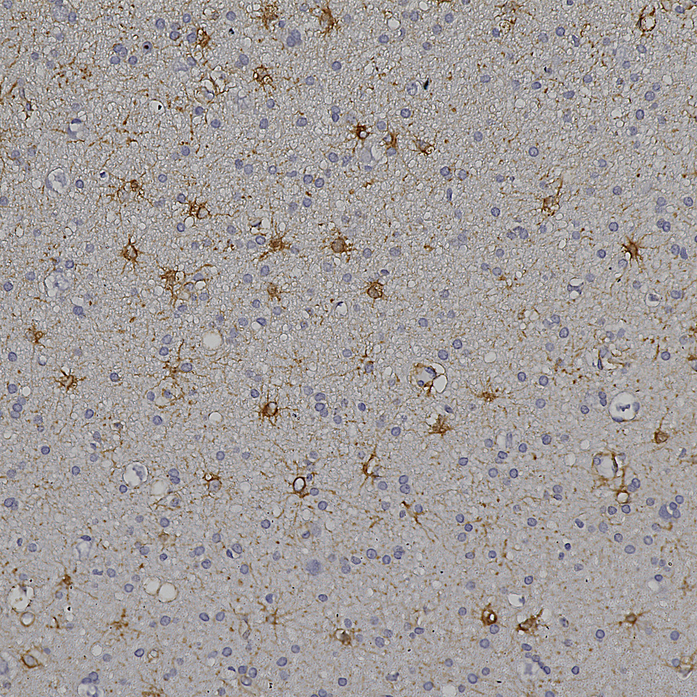
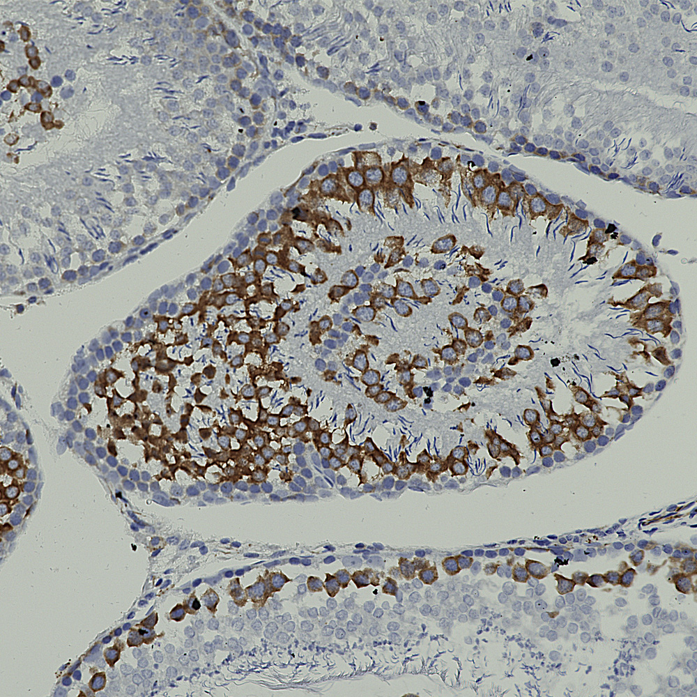
And below is an example of IHC of rat testis stained with our antibody to Nestin, MCA-4D11.
Below is rat hippocampus stained with MCA-4H5 one of our MAP2 antibodies showing staining for hippocampal pyramidal cells.
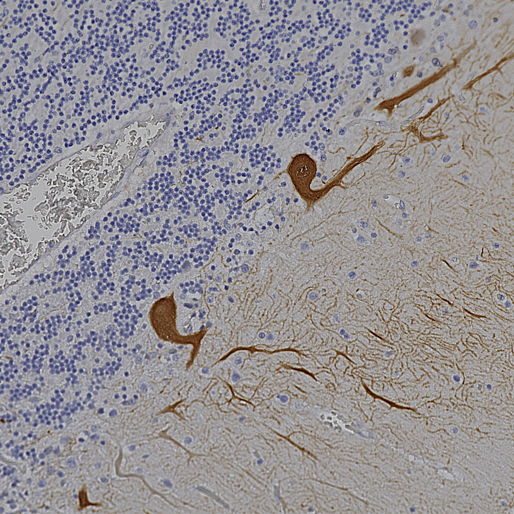
Below is an IHC specimen of human cerebellum stained with MCA-4H7 mouse monoclonal antibody to calbindin which reveals Purkinke cell dendrites in the molecular layer.
Below is an example of IHC staining with MCA-5B10, our epitope mapped monoclonal antibody to MAP-τ showing flame shaped tangles in the hippocampus of an Alzheimer’s disease patient.
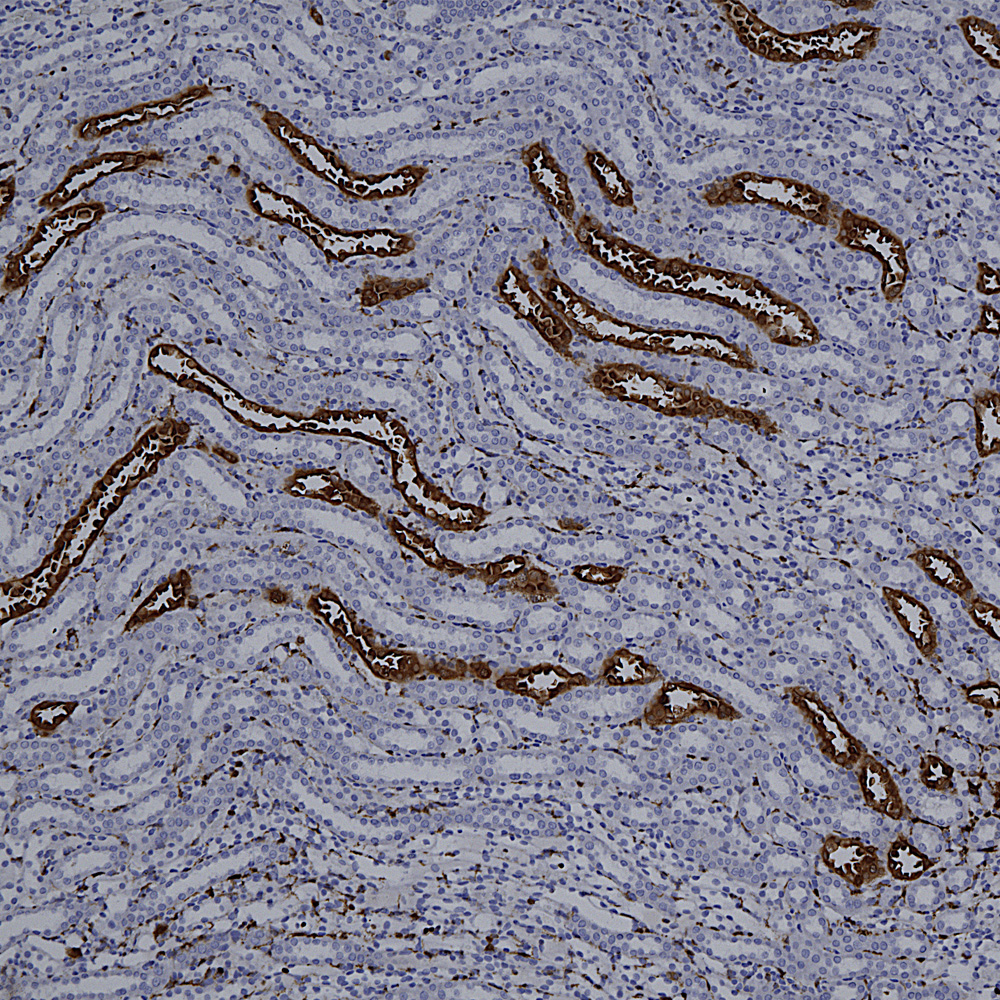
Below is a section of rat kidney stained with MCA-5C21, our monoclonal antibody to Galectin 3.
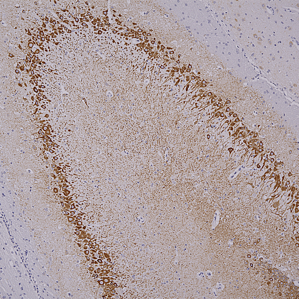
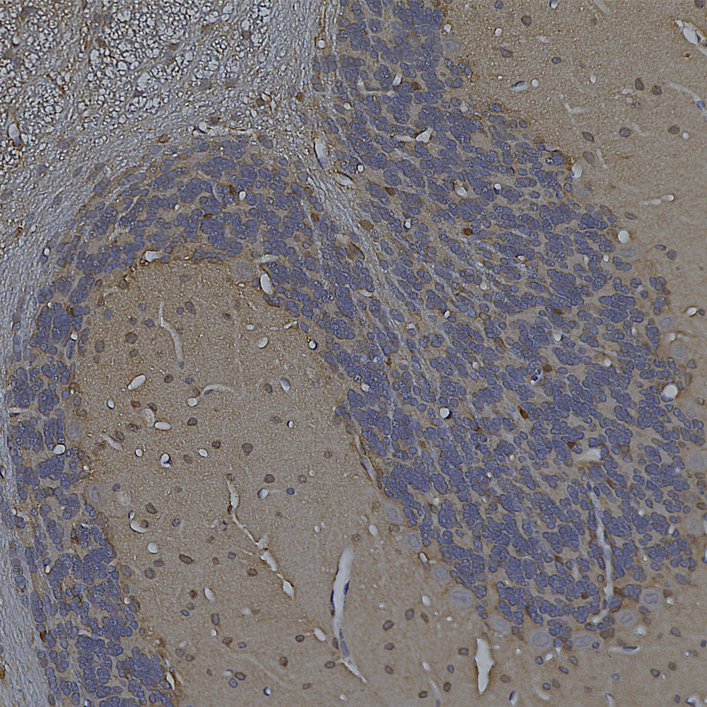
Below is IHC of rat cerebellum stained with our monoclonal antibody to MAP2 MCA-5H11, showing prominent Purkinje cell bodies and dendrites in the molecular layer.
Finally here is IHC of rat cerebellum stained with our MCA-253 monoclonal to non-neuronal enolase, also known as Eno1 or α-enolase, which stains astrocytes and other non-neuronal cells.
March 2023 News
The data in our recent publication in BioRχiv preprint, entitled “Uman Type NF-L Antibodies Are Effective Reagents for the Imaging of Neurodegeneration” was edited and modified to include further data and was submitted for peer review at the journal “Brain Communications“. It has now been accepted for publication and an unedited and unformatted version of the paper appeared on-line, see 10.1093/braincomms/fcad067. The same link will access the edited and formatted version of the paper which will be available soon.
We also add new data for many of our mouse monoclonal antibodies which we have now screen for immunohistochemistry (IHC) on paraffin embedded and formalin fixed human and rodent tissues. For example our widely used mouse monoclonal antibody to c-FOS, MCA-2H2, works well on rat hippocampus highlighting a few active neurons. These cells were expressing large amounts of c-FOS and therefore activating transcription of other genes at the time the tissue was processed and with current techniques can be characterized in terms of mRNA expression and connections. This data is shown under the “Additional Info” tag on the MCA-2H2 web page. Many of our antibodies perform very well and we have posted images under the “Additional Info” tag on the relevant web pages. As expected a few antibodies did not work well and we have added “not recommended for IHC” on the web page. We are currently screening our entire panel of mouse monoclonal antibodies for utility in IHC, and will shortly post results of this.