June 2023 News
We have now finished testing all our rabbit polyclonal antibodies for immunohistochemistry on formalin fixed and paraffin embedded (FFPE) rodent and rodent tissues. So we found that many of them work very well, as shown in the examples below. So now we can recommend (or not) all of our antibodies on FFPE human and rodent specimens, describe the protocols we used and show convincing example images.
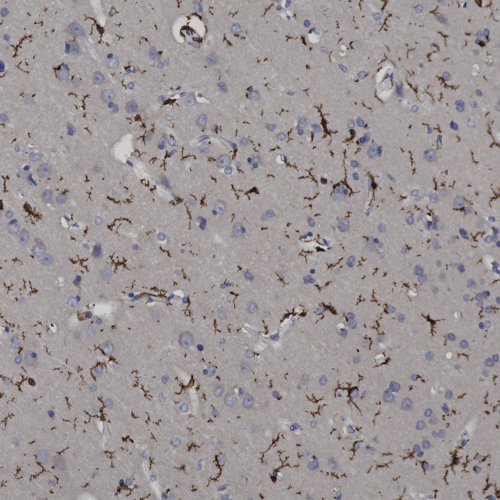
Chromogenic immunostaining of a NBF fixed paraffin embedded human brain cortex section with rabbit pAb to IBA1, RPCA-IBA1, dilution 1:2,000, detected with DAB (brown) using the Vector Labs ImmPRESS method and reagents with citra buffer retrieval. Hematoxylin (blue) was used as the counterstain. The RPCA-IBA1 antibody specifically labels the cytoplasm of microglial cells. This antibody performs well in testing with 4% PFA and standard NBF fixed mouse, rat and human tissue.
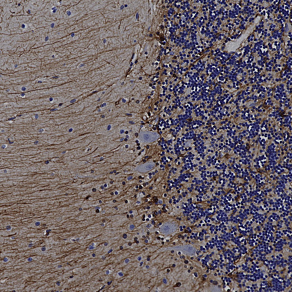
Chromogenic immunostaining of a formalin fixed paraffin embedded human cerebellum section with rabbit pAb to GFAP, RPCA-GFAP, dilution 1:5,000, detected with DAB (brown) using the Vector Labs ImmPRESS method and reagents with citra buffer retrieval. Hematoxylin (blue) was used as the counterstain. The RPCA-GFAP antibody labels the processes of astrocytes within both the granular and molecular layers and Bergmann glia in the molecular layer. This antibody performs well in testing with both 4% PFA and standard NBF fixed rat, mouse, and human tissues.
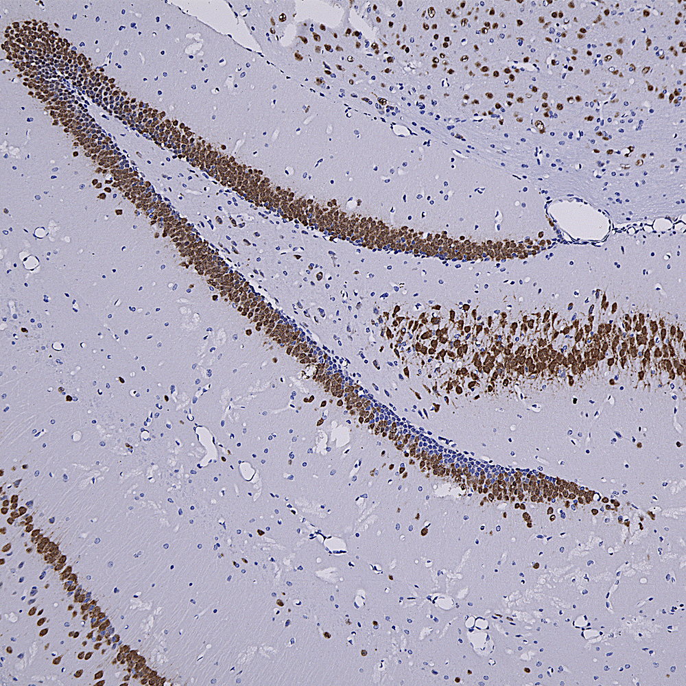
Chromogenic immunostaining of a formalin fixed paraffin embedded mouse hippocampus section with rabbit pAb to Fox3/NeuN, RPCA-FOX3, dilution 1:4,000, detected with DAB (brown) using the Vector Labs ImmPRESS method and reagents with citra buffer retrieval. The RPCA-FOX3 antibody selectively labels the nuclei and distal perikaryal of most neuronal cell populations. This antibody performs well in testing with 4% PFA and NBF fixed mouse, human, and rat tissues. Mouse select image for larger view.
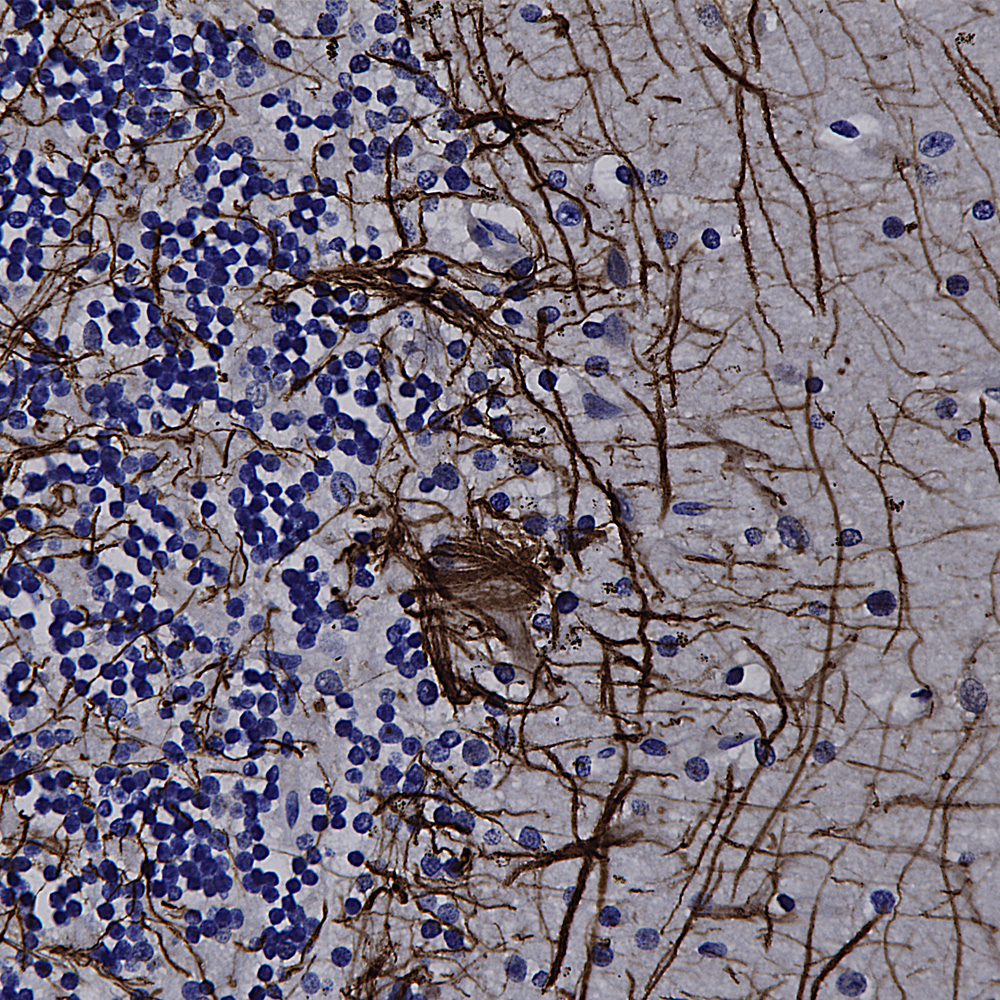
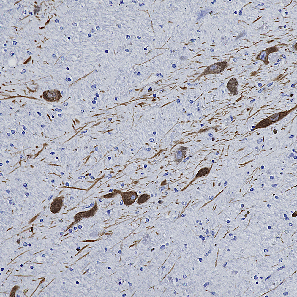
Chromogenic immunostaining of a formalin fixed paraffin embedded human cerebellum section with rabbit pAb to NF-L, RPCA-NF-L-ct, dilution 1:5,000, detected with DAB (brown) using the Vector Labs ImmPRESS method and reagents with citra buffer retrieval. Hematoxylin (blue) was used as the counterstain. RPCA-NF-L-ct strongly labels the axons and dendrites of Purkinje cells and the projections of neuronal cells within the granular layer. This antibody performs well in testing with both 4% PFA and standard NBF fixed rat, mouse, and human tissues. Mouse select image for larger view.
Chromogenic immunostaining of a NBF fixed paraffin embedded human midbrain section with rabbit pAb to tyrosine hydroxylase, RPCA-TH, dilution 1:10,000, detected with DAB (brown) using the Vector Labs ImmPRESS method and reagents with citra buffer retrieval. Hematoxylin (blue) was used as the counterstain. In this image, RPCA-TH antibody labels dopaminergic neurons and their axons traversing the striatum. This antibody performs well in testing with 4% PFA and standard NBF fixed mouse, rat and human tissue. Mouse select image for larger view.