We have fruitful and ongoing interactions with other well regarded companies such as Neuroscience Associates and LifeCanvas Technologies. Neuroscience Associates are a long standing company located in Knoxville, Tennessee which specializes in tissue sectioning and staining. We provide them with antibodies and certain custom products. Lifecanvas is a newer company located in Cambridge, Massachusetts, which specializes in cutting edge tissue “clearing” methods. These methods allow tissue specimens to be rendered transparent so that flourescent signals, either from fluorescent proteins or from antibody methods, can be documented in thick specimens. This allows for the efficient documentation of 3D structures. For one example see here:
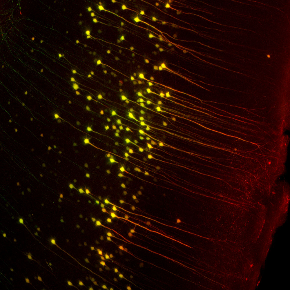
Above, mice transgenic for green fluorescent protein (GFP) under the Thy1 promoter express GFP in cerebral pyramidal cells. The image is of a 400μM maximum intensity optical section taken from an entire Thy1-GFP mouse brain “cleared” in the Lifecanvas lab. Lifecanvas scientists then stained the brain with EnCor goat polyclonal antibody to GFP GPCA-GFP coupled to a red dye. As expected, the green and red signals superimpose in the perikarya of the pyramidal cells which appear yellowish. The antibody also stains the apical dendrites which have a lower level of GFP expression, effectively amplifying the GFP signal. Mouse select on image for larger view.
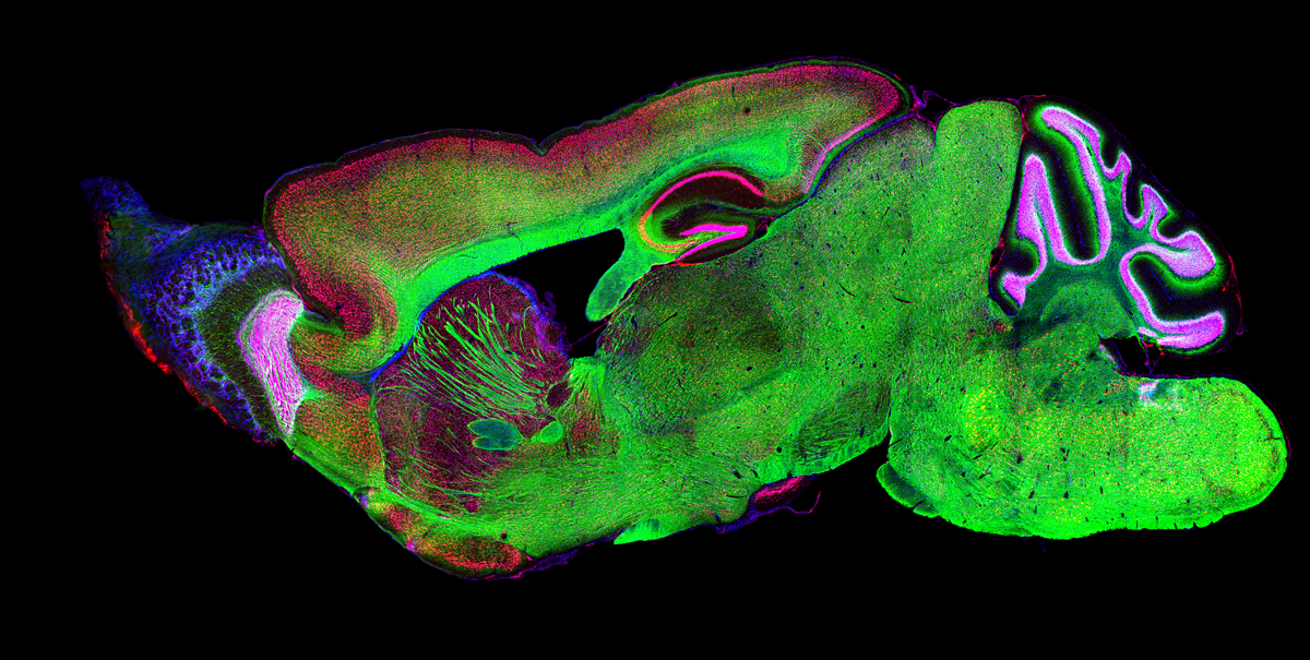
Above, sagittal section of mouse brain stained with mouse monoclonal antibody to Fox3/NeuN MCA-1B7 in red and rabbit polyclonal antibody to the C-terminus of neurofilament NF-L RPCA-NF-L-ct in green. Nuclear DNA is revealed by DAPI in blue. The FOX3/NeuN antibody reveals neuronal nuclei and cell bodies while the NF-L antibody binds to the core structure of axons. DNA, revealed in blue with the DAPI stain, is found in the nuclei of all types of brain cell. Mouse select on image for larger view.
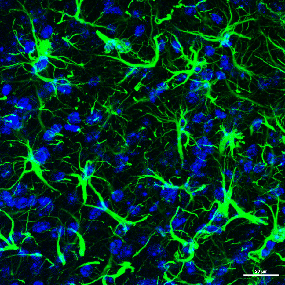
We have many users of our immunoreagents in the University of Florida. Here is an image made by Dr. Fatemeh Shaerzadeh, working in the lab of Professor Habibeh Khoshbouei in the McKnight Brain Institute. A section of mouse cerebral cortex was stained with EnCor chicken polyclonal antibody to GFAP CPCA-GFAP in green and DNA in blue. The fibrous component of the processes of astroctyes are revealed. Mouse select on image for larger view.
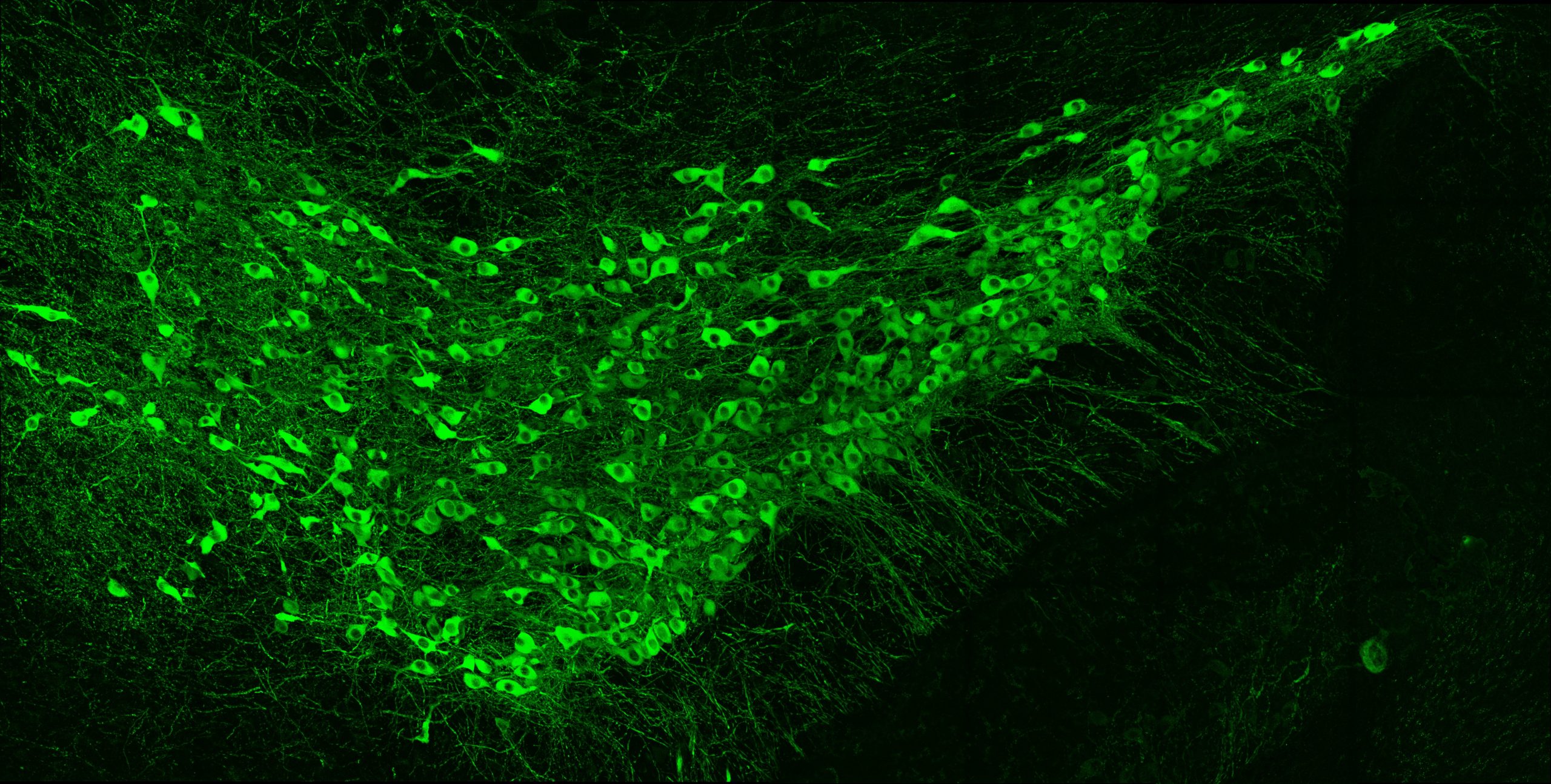
Above is an image made by Dr. Fatimeh Shaerzadeh, working in the lab of Professor Habibeh Khoshbouei in the McKnight Brain Institute of the University of Florida. A section of mouse midbrain stained with EnCor mouse monoclonal antibody to tyrosine hydroxylase MCA-4H2 in green. The cytoplasm and processes of these dopaminergic neurons are revealed. Mouse select on image for larger view.
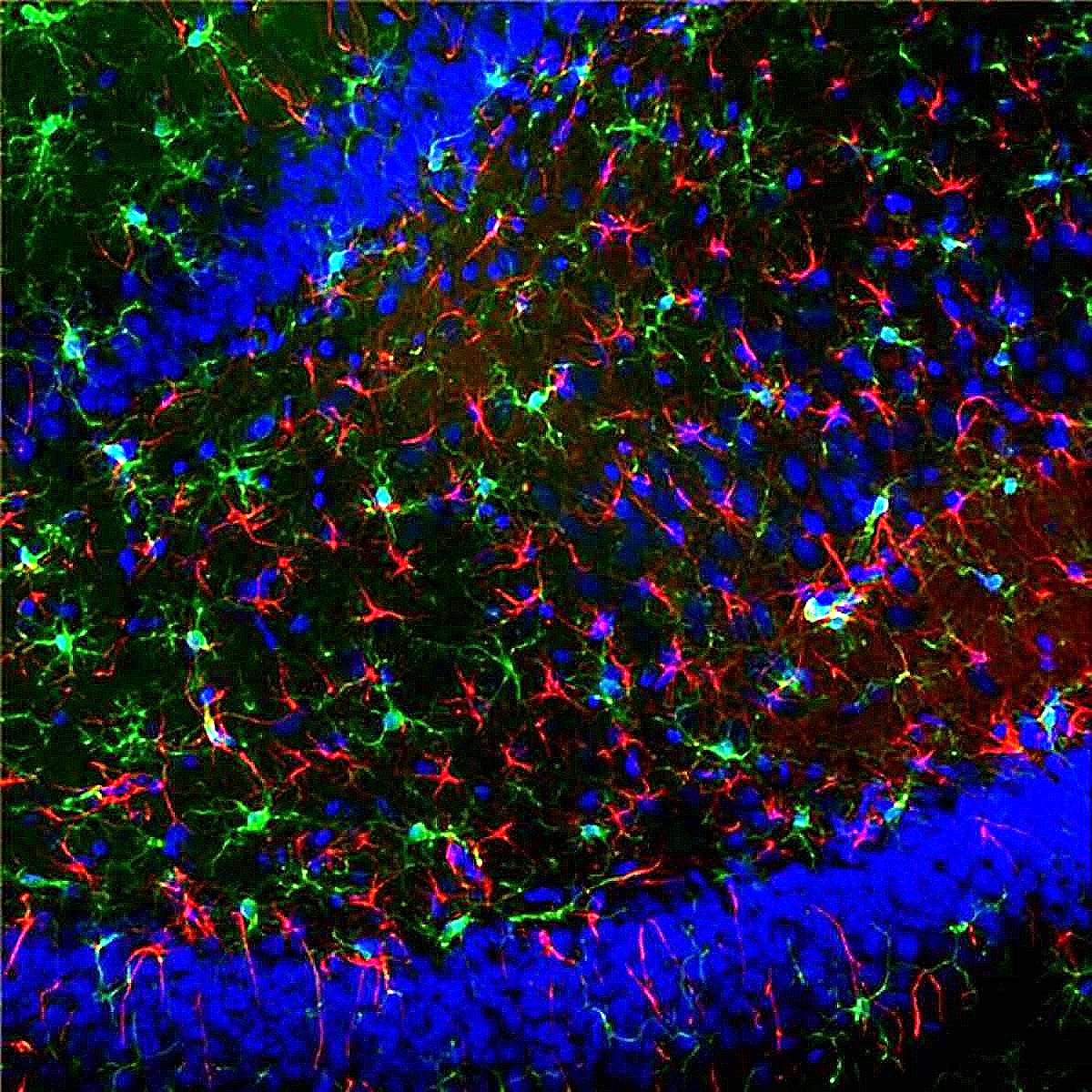
The image is made by researchers Fatimah Ahmad, Hajar Benmhammed and Vera Dermesrobian, in the lab of Dr. Firas Kobeissy at the American University of Beirut. The image shows a section of female rat dentate gyrus. Star-shaped astrocytes are stained with EnCor chicken polyclonal antibody to GFAP, CPCA-GFAP in red, and microglia are stained with antibody to Iba1 in green. DNA is revealed usinf DAPI stain in blue. Mouse select on image for larger view.