

EnCor Biotechnology
Mouse Monoclonal Antibody to Aldehyde Dehydrogenase 1 Family Member L1 (ALDH1L1), Cat# MCA-2E7
Description
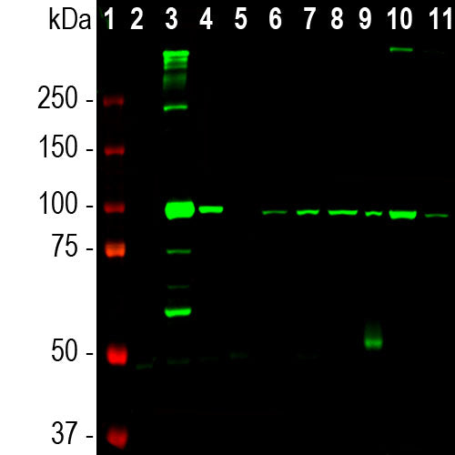
The MCA-2E7 antibody was made against amino acids 402-902 of human ALDH1L1 protein expressed in and purified from E. coli. It works for western blots and IHC on human and rodent material, but is not recommended for ICC of floating sections or on overly fixed IHC tissue. We market an alternate mouse monoclonal to ALDH1L1, MCA-4A12 as well as a rabbit polyclonal antibody, RPCA-ALDH1L1. ALDH1L1 antibodies are useful markers for the identification of astrocytic cells.
Add a short description for this tabbed section
| Immunogen: | Amino acids 402-902 of human ALDH1L1 protein expressed in and purified from E. coli. |
| HGNC Name: | ALDH1L1 |
| UniProt: | O75891 |
| Molecular Weight: | 100kDa |
| Host: | Mouse |
| Isotype: | IgG1 |
| Species Cross-Reactivity: | Human, rat, mouse |
| RRID: | AB_2572220 |
| Format: | Protein G affinity purified antibody at 1mg/mL in 50% PBS, 50% glycerol plus 5mM NaN3 |
| Applications: | WB, ICC, IHC(p) |
| Recommended Dilutions: | WB: 1:5,000-1:10,000. ICC: 1:1,000. IHC: 1:1,000. |
| Storage: | Store at 4°C for short term, for longer term store at -20°C. Stable for 12 months from date of receipt. |
Aldehyde dehydrogenase family 1, member L1 (ALDH1L1) is a cytosolic enzyme and one member of a large family of aldehyde dehydrogenases. ALDH1L1 catalyses the NADP(+) dependent oxidation of 10-formyltetrahydrofolate to tetrahydrofolate and Carbon dioxide (1). ALDH1L1 expression is highly tissue specific, with very high levels in the liver, representing up to 1% of the total pool of soluble cell proteins. Cahoy et al. used fluorescent activated cell sorting to isolate astrocytes from enhanced green fluorescent protein (GFP) expressing transgenic mice, with GFP expression being under the control of the S100β promoter, expected to direct GFP to astrocytes. They then created a transcriptome database of the gene expression levels using Affymetrix GeneChip arrays (2). They identified ALDH1L1 mRNA as very abundant and expressed only in astrocytes, suggesting that ALDH1L1 protein would be a expressed at high levels and only in astrocytes. Based on immunocytochemical studies they claimed that ALDH1L1 is more widely expressed in astrocytes throughout the brain, while the widely used astrocyte marker GFAP shows more predominant expression in white matter. The also claimed that AlDH1L1 expression gives a more detailed view of astrocyte morphology since it is expressed throughout the cell including fine protoplasmic protrusions. In contrast GFAP is found in the intermediate filament core of the astrocyte, and these filaments are not found in finer cytoplasmic protrusions. Loss of function or expression of ALDH1L1 is associated with decreased apoptosis, increased cell motility, and cancer progression, suggesting its role as a potential biomarker and a target in cancer therapy (3-5).

Chromogenic immunostaining of a 4% PFA fixed paraffin embedded rat cerebellum section with mouse mAb to ALDHL1L, MCA-2E7, dilution 1:1,000, detected with DAB (brown) using the Vector Labs ImmPRESS method and reagents with citrate buffer retrieval. Hematoxylin (blue) was used as the counterstain. This antibody performs well in testing with both 4% PFA and standard NBF fixed tissues. Mouse select image for larger view.
1. Kisliuk RL. Folate biochemistry in relation to antifolate selectivity. In Jackman AL, editor. Antifolate drugs in cancer therapy. Humana Press 13-36 (1999).
2. Cahoy JD, et al. A transcriptome database for astrocytes, neurons, and oligodendrocytes: a new resource for understanding brain development and function. J. Neurosci. 28:264-78 (2008).
3. Krupenko SA, Oleinik NV. 10-formyltetrahydrofolate dehydrogenase, one of the major folate enzymes, is down-regulated in tumor tissues and possesses suppressor effects on cancer cells. Cell Growth Differ. 13:227-36 (2002).
4. Rodriguez FJ, et al. Gene expression profiling of NF-1-associated and sporadic pilocytic astrocytoma identifies aldehyde dehydrogenase 1 family member L1 (ALDH1L1) as an underexpressed candidate biomarker in aggressive subtypes. J. Neuropath. Exp. Neurol. 67:1194-204 (2008).
5. Oleinik NV, Krupenko NI, Krupenko SA. Epigenetic Silencing of ALDH1L1, a Metabolic Regulator of Cellular Proliferation, in Cancers. Genes Cancer 2:130-9 (2011).
Add a short description for this tabbed section


,%20Cat%23%20MCA-2E7)