

EnCor Biotechnology
Mouse Monoclonal Antibody to Phosphoprotein Enriched in Astrocytes (PEA-15), Cat# MCA-4D117
Description
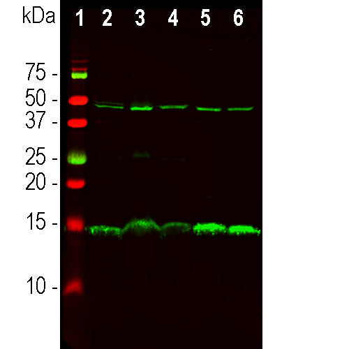
MCA-4D117 was made against a recombinant full length PEA-15 construct expressed in and purified from E. coli. The antibody can be used for western blotting IF, ICC and IHC (see "Additional Data" tab). The antibody highlights a subset of cell bodies in CNS tissues.
Add a short description for this tabbed section
| Immunogen: | Full length recombinant human PEA-15 as expressed in and purified from E. coli. |
| HGNC Name: | PEA-15 |
| UniProt: | Q15121 |
| Molecular Weight: | 15kDa |
| Host: | Mouse |
| Isotype: | IgG1 |
| Species Cross-Reactivity: | Human, Rat, Mouse, Cow |
| RRID: | AB_2861183 |
| Format: | Protein G affinity purified antibody at 1mg/mL in 50% PBS, 50% glycerol plus 5mM NaN3 |
| Applications: | WB, ICC/IF, IHC |
| Recommended Dilutions: | WB: 1:1,000-1:2,000. ICC/IF, and IHC: 1:500-1:1,000. |
| Storage: | Store at 4°C for short term, for longer term store at -20°C. Stable for 12 months from date of receipt. |
Pea15 was originally isolated as a major low molecular weight of embryonic mouse striatal astrocytes grown in cell culture. Three spots on 2D gels with an apparent molecular weight of 15kDa and isoelectric point 5.1-5.3 were shown to be different forms of one protein. The protein was serine phosphorylated on one site by protein kinase C both in vivo and in vitro and the protein was named "phosphoprotein enriched in astrocytes of 15kDa", hence PEA-15 (1). Subsequent cloning and sequencing revealed a protein well conserved in sequence between mouse and human and which was heavily expressed in brain (2). Independently the same protein was found to be upregulated in fibroblasts and tissues of diabetic patients, and has hence named "protein enriched in diabetes" or PED (3). Immunocytochemical studies showed that the protein was heavily expressed in astrocytes and certain neurons in the CNS of mice, though it is expressed a lower levels ubiquitously (2,4). The protein could be phosphorylated on a second site by either CaMKII or Akt/PKB, and further examination of the amino acid sequence revealed an N-terminal death effector domain (DED), a predominantly α-helical domain found in many proteins which function in the regulation of apoptosis (5). PEA-15 was shown to interact with extracellular signal regulated kinase (ERK, 6) and regulate the nuclear entry of this protein, and several other important interactions with other proteins involved in regulation of apoptosis, glucose metabolism and cell growth have been described (reviewed in 7).

Chromogenic immunostaining of a 4% PFA fixed paraffin embedded rat cerebellum section with mouse mAb to PEA-15, MCA-4D117, dilution 1:1,000, detected with DAB (brown) following the the Vector Labs ImmPRESS method and reagents with citrate buffer retrieval. Hematoxylin (blue) was used as the counterstain. PEA-15 stains the cytoplasm of subpopulations of neurons and astrocytes throughout the brain. This antibody performs well in testing with 4% PFA but does not stain long term NBF fixed tissue effectively. Mouse select image for larger view.

Chromogenic immunostaining of a 4% PFA fixed paraffin embedded rat testes section with mouse mAb to PEA-15, MCA-4D117, dilution 1:1,000, detected with DAB (brown) following the the Vector Labs ImmPRESS method and reagents with citrate buffer retrieval. Hematoxylin (blue) was used as the counterstain. Mouse select image for larger view.
1. Araujo, H. et al. Characterization of PEA-15, a major substrate for protein kinase C in astrocytes. J. Biol. Chem. 268:5911-20 (1993).
2. Estelles, A. et al. The Major Astrocytic Phosphoprotein PEA-15 Is Encoded by Two mRNAs Conserved on Their Full Length in Mouse and Human. J. Biol. Chem. 271:14800-6 (1996).
3. Condorelli, G. et al. PED/PEA-15 gene controls glucose transport and is overexpressed in type 2 diabetes mellitusEMBO J. 17:3858-66 (1998).
4. Sharif, A. et al. The expression of PEA-15 (phosphoprotein enriched in astrocytes of 15 kDa) defines subpopulations of astrocytes and neurons throughout the adult mouse brain. Neurosci. 126:263-75 (2004).
5. Boldin, MP. et al. A novel protein that interacts with the death domain of Fas/APO1 contains a sequence motif related to the death domain. J. Biol. Chem. 271:7795–78 (1995).
6. Ramos, JW. et al. PEA-15 Mediates Cytoplasmic Sequestration of ERK MAP Kinase. Dev. Cell 1:239–50 (2001).
7. Fiory, F. et al. Frontiers: PED/PEA-15, a multifunctional protein controlling cell survival and glucose metabolism. Am. J. Physiol. Endocrinol. Metab. 297:E592-601 (2009).
Add a short description for this tabbed section


,%20Cat%23%20MCA-4D117)