

EnCor Biotechnology
Mouse Monoclonal Antibody to β-Tubulin, Cat# MCA-4E4
Description
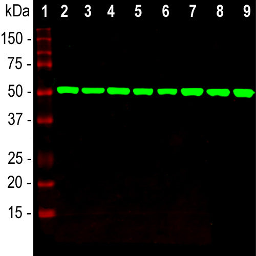
The MCA-4E4 antibody was raised against tubulin purified from pig brain and reacts with recombinant β-tubulin (Abcam), but not recombinant α-tubulin (Abnova) by ELISA and dot blots. β-tubulin is regarded as a “house keeping” protein which is generally not altered much in expression as a result of experimental manipulations. As a result antibodies to β-tubulin are widely used as loading controls in western blotting experiments as a standard by which the levels of other proteins may be measured. As shown here, MCA-4E4 produces a single strong clean band on homogenates of cell and tissue extracts. It also produces beautiful images of the microtubular network of cells grown in culture and can be used in IHC staining of tissue sections. MCA-4E4 is an IgG2a class antibody and an alternate antibody useful for certain kinds of experiment is MCA-1B12, an antibody of similar β-tubulin specificity.
Add a short description for this tabbed section
| Immunogen: | Pig brain tubulin preparation |
| HGNC Name: | TUBB |
| UniProt: | P02554 |
| Molecular Weight: | 50kDa |
| Host: | Mouse |
| Isotype: | IgG2a |
| Species Cross-Reactivity: | Human, Monkey, Rat, Mouse |
| RRID: | AB_2492290 |
| Format: | Protein G affinity purified antibody at 1mg/mL in 50% PBS, 50% glycerol plus 5mM NaN3 |
| Applications: | WB, ICC, IHC |
| Recommended Dilutions: | WB: 1:5,000-1:10,000. ICC/IF: 1:2000-1:5,000. IHC: 1:1,000-1:2,000. |
| Storage: | Store at 4°C for short term, for longer term store at -20°C. Stable for 12 months from date of receipt. |
Tubulins are a major class of cytoskeletal proteins and divided into five distinct classes, namely α, β, γ, δ and ε. The most abundant members of this family are the α and β-tubulins which are the major components of cytoplasmic microtubules. The various subunits have molecular weights of approximately 50kDa and are 50% identical to one another at the protein sequence level. Microtubules are assembled from stable dimers of one α and one β subunit, and regulated polymerization and depolymerization of these dimers controls the number and location of microtubules in cells (1,2). Microtubules are involved in a number of essential cellular functions including the maintenance of cell shape, vesicle and organelle transport, cell motility, cell signaling, meiosis and mitosis. The important role of microtubules in forming the mitotic spindle during cell division makes them a desirable target for the development of therapeutic agents directed against rapidly dividing cancer cells (3). For example, Taxol, a.k.a. Paclitaxel, is a low molecular weight drug which binds αβ tubulin dimers and prevents their polymerization. This prevents formation of the mitotic spindle, inhibits cell division and so halts tumor growth. For an interesting review of the first 50 years of tubulin research see reference 4

Chromogenic immunostaining of a 4% PFA fixed paraffin embedded rat cerebral cortex section with mouse mAb to β-tubulin, MCA-4E4, dilution 1:1,000, detected with DAB (brown) using the Vector Elite ABC-HRP detection and reagents with citrate buffer retrieval. Hematoxylin (blue) was used as the counterstain. MCA-4E4 antibody labels the cytoplasm of neurons (pyramidal cells in this image) and endothelial cells as well as the neuropil. This antibody performs well in testing with both 4% PFA and standard NBF fixed tissues. Mouse select image for larger view.
1. Nogales E, Wolf SG, Downing KH. Structure of the alpha-beta tubulin dimer by electron crystallography. Nature 391:199-203 (1998)..
2. Nogales E. Structural insight into microtubule function. Ann. Rev. Biophys. Biomol. Struct. 30:397-420 (2001).
3. Perez EA. Microtubule inhibitors: Differentiating tubulin-inhibiting agents based on mechanisms of action, clinical activity, and resistance. Mol. Cancer Ther. 8:2086-95 (2009).
4. Borisy G, et al. Microtubules: 50 years on from the discovery of tubulin. Nat. Rev. Mol. Cell Biol. 17:322-8 (2016).
Add a short description for this tabbed section


