
EnCor Biotechnology
Mouse Monoclonal Antibody to Lamin A/C, Cat# MCA-4C4
Description
The MCA-4C4 was raised against full length recombinant human lamin A, and binds human lamin C also. However it does not recognize rodent lamin A or C. Since MCA-4C4 antibody is human specific, it can be used to monitor human material in a rodent background. It works well on western blots and for IF and ICC but is not recommended for IHC. We also market a chicken polyclonal antibody to lamin A/C CPCA-LaminAC with similar properties to MCA-4C4.
Add a short description for this tabbed section
| Immunogen: | Full length recombinant human lamin A expressed in and purified from E. coli. |
| HGNC Name: | LMNA |
| UniProt: | P02545 |
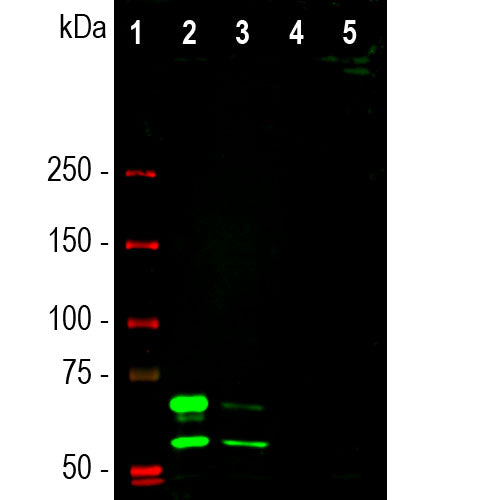
| Molecular Weight: | 65kDa, 74kDa |
| Host: | Mouse |
| Isotype: | IgG1 |
| Species Cross-Reactivity: | Human, unreactive with rodent |
| RRID: | AB_2572339 |
| Format: | Protein G affinity purified antibody at 1mg/mL in 50% PBS, 50% glycerol plus 5mM NaN3 |
| Applications: | WB, IF/ICC |
| Recommended Dilutions: | WB: 1:1,000-1:2,000. IF/ICC: 1:1,000. IHC: Not Recommended. |
| Storage: | Store at 4°C for short term, for longer term store at -20°C. Stable for 12 months from date of receipt. |
Lamin A and lamin C are members of the intermediate filament protein family and are located in the nucleus where they function as skeletal components of the inner nuclear membrane (1). The two proteins are generated by alternate transcription from the single LMNA gene. Lamin A has a molecular weight of about 74kDa while lamin C is 65kDa. The lamin A protein includes a C-terminal segment of 98 amino acids missing from lamin C, while lamin C has a unique C-terminal 6 amino acid peptide not present in lamin A. As a result antibodies raised against lamin A are very likely bind to lamin C, as is the case with MCA-4C4. During cell division the nuclear lamina breaks down and lamin A/C containing filaments depolymerize, this being regulated by phosphorylation by cyclin dependent protein kinase 1 (CDK1). Mutations in the lamin A/C gene are associated with several serious human diseases, including Emery-Dreifuss muscular dystrophy, familial partial lipodystrophy, limb girdle muscular dystrophy, dilated cardiomyopathy, Charcot-Marie-Tooth disease type 2B1, Hutchinson-Gilford progeria syndrome and Hutchinson-Gilford progeria syndrome (3-6).
This antibody has been tested on formalin fixed and paraffin embedded samples for IHC, and is not recommended for this purpose.
1. Fisher DZ, Chaudhary N, Blobel G. cDNA sequencing of nuclear lamins A and C reveals primary and secondary structural homology to intermediate filament proteins. PNAS 83:6450-54 (1986).
2. McKeon FD, Kirschner MW, Caput D. Homologies in both primary and secondary structure between nuclear envelope and intermediate filament proteins. Nature 319: 463-8 (1986).
3. Bonne G, et al. Mutations in the gene encoding lamin A/C cause autosomal dominant Emery-Dreifuss muscular dystrophy. Nat. Genet. 21:285-8 (1999).
4. Novelli G, et al. Mandibuloacral dysplasia is caused by a mutation in LMNA-encoding lamin A/C. Am. J. Hum. Genet. 71:426-31 (2002).
5. De Sandre-Giovannoli A, et al. Homozygous Defects In Lmna, Encoding Lamin A/C Nuclear‐Envelope Proteins, Cause Autosomal Recessive Axonal Neuropathy In Human (Charcot‐Marie‐Tooth Disorder Type 2) And Mouse Am. J. Hum. Genet. 70:726-36 (2002).
6. Liu B and Zhou Z. Lamin A/C, laminopathies and premature ageing. Histol. Histopathol. 23:747-63 (2006).
Add a short description for this tabbed section


