

EnCor Biotechnology
Mouse Monoclonal Antibody to Glial Fibrillary Acidic Protein (GFAP), Cat# MCA-5C10
Description
The MCA-5C10 antibody was made against purified GFAP from porcine spinal cord, EnCor product PROT-m-GFAP. High quality antibodies to GFAP such as MCA-5C10 are useful for visualizing glia and monitoring developmental, disease and damage related CNS alterations. This antibody has been shown to work well on western blots, IF, ICC and IHC and also to recognize GFAP from a variety of species. The epitope is dependent on sequence from 257-276, a region including a caspase 3 cleavage site. Interestingly the MCA-5C10 epitope is destroyed during injury induced degeneration. We also supply alternate mouse monoclonal antibodies, MCA-2A5 and MCA-3E10, which are particularly useful for human studies. We also developed chicken, rabbit and goat polyclonals, CPCA-GFAP, RPCA-GFAP, and GPCA-GFAP respectively.
- Cell Structure Marker
- Cell Type Marker
- Cytoskeletal Marker
- Developmental Marker
- Epitope Mapped Antibodies
- Immunohistochemistry Verified
- Mouse Monoclonal Antibodies
- Pathology Related Marker
Add a short description for this tabbed section
| Immunogen: | GFAP purified from porcine spinal cord |
| HGNC Name: | GFAP |
| UniProt: | P14136 |
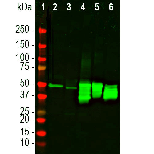
| Molecular Weight: | 50kDa |
| Host: | Mouse |
| Isotype: | IgG1 heavy, κ light |
| Species Cross-Reactivity: | Human, Rat, Mouse, Cow, Pig, Horse |
| RRID: | AB_2572311 |
| Format: | Protein G affinity purified antibody at 1mg/mL in 50% PBS, 50% glycerol plus 5mM NaN3 |
| Applications: | WB, IF/ICC, IHC |
| Recommended Dilutions: | WB: 1:5,000. IF/ICC, or IHC: 1:1,000-1:2,000. |
| Storage: | Store at 4°C for short term, for longer term store at -20°C. Stable for 12 months from date of receipt. |
Glial fibrillary acidic protein (GFAP) is strongly and specifically expressed in astrocytes, Bergmann glia, certain other glia in the central nervous system, in satellite cells in peripheral ganglia, and in non-myelinating Schwann cells in peripheral nerves. GFAP expression is also seen in developing neural stem cells and GFAP levels may greatly increase in regions of CNS injury or disease. The formation of a GFAP rich "glial scar" following CNS injury may be one reason why reconnection of severed processes is relatively inefficient in adults. Point mutations in the GFAP gene are causative of Alexander disease (5). All forms of Alexander disease are characterized by the presence of Rosenthal fibers, which are GFAP containing cytoplasmic inclusions found in astrocytes. Some interest has recently been focused on GFAP as a protein released into blood and CSF following traumatic brain injury, stroke and other CNS compromises (6,7). Measurement of the levels of blood or CSF GFAP may give information about patient presentation, progress, response to therapy or outcome.

Chromogenic immunostaining of a formalin fixed paraffin embedded human cerebellum stained with mouse mAb, MCA-5C10, dilution 1:2,000, detected with DAB (brown) using the the Vector Labs ImmPRESS method and reagents with citrate buffer retrieval. Hemotoxylin (blue) was used as the counter. Mouse select image for larger view.

This is one of a series of formalin fixed paraffin embedded adult horse brain samples generated by Maureen T. Long in the Department of Infectious Diseases and Pathology, in the College of Veterinary Medicine, University of Florida. The findings were recently published in the Equine Veterinary Journal, link here. The samples were fixed and embedded in paraffin in 2007 and sections were kept at room temperature until 2011. The sections were processed for antigen retrieval by boiling in pH=6 Citrate buffer for 10 min. Primary incubation with MCA-5C10 was for 1 hour at 37°C, and secondary antibody incubation and color reaction was performed using the Vector mouse ABC kit. Mouse select image for larger view.

Section of rat cerebellum processed using our standard ICC on floating sections with our mAb to GFAP MCA-5C10, 1:2,000 dilution. The image shows Bergmann radial glial sending processes to the pia in the molecular layer. The granular layer is revealed with the DAPI DNA dye which highlights the nuclei of granule neurons. The granular and central white matter contain astrocytes which can be seen sending GFAP positive processes and end feet out to the blood vessels.

Rat cerebellum stained with mAb MCA-5C10 1:2,000 showing the molecular layer at the left and the granular layer at the right. The processes of the Bergmann radial glia are prominent in the molecular layer while the processes of astrocytes are seem in the molecular layer. The sample was processed using our standard IHC protocol with DAB (brown) using the the Vector Labs ImmPRESS method and reagents with citrate buffer retrieval. Section was lightly counterstained with hemotoxylin. Mouse select for larger view.

Section of human cerebellum processed with MCA-5C10 showing molecular layer to the left and the granular layer at the edge to the right. The processes of Bergmann glia can be see in the molecular layer and the processes of astrocytes in the granular layer. The sample was processed using our standard IHC protocol with DAB (brown) using the the Vector Labs ImmPRESS method and reagents with citrate buffer retrieval. Section was lightly counterstained with hemotoxylin. Mouse ellect for larger view.

Immunofluorescent analysis of mouse brain stained with mouse mAb to GFAP, MCA-5C10, dilution 1:5,000 in green, and costained with rabbit pAb to FOX3/NeuN, RPCA-FOX3, dilution 1:2,000 in red. Blue is Hoechst staining of nuclear DNA. Following transcardial perfusion of mouse with 4% paraformaldehyde, brain was post fixed for 24 hours, cut to 40μM, and free-floating sections were stained with above antibodies. The MCA-5C10 antibody stains processes of astrocytic glial cells, while the RPCA-FOX3 antibody labels nuclei and cytoplasm of certain neurons. Since mouse tissues contain mouse IgGs notably in blood vessels, using a mouse antibody on mouse tissue is not recommended. We can recommend our rabbit or chicken polyclonal antibodies to GFAP RPCA-GFAP or CPCA-GFAP for this purpose.
This antibody is widely sold through EnCor OEM partners, and a growing list of peer-reviewed publications cite EnCor as origin of this antibody, see here.
1. Bignami A, Eng LF, Dahl D, Uyeda CT. Localization of the glial fibrillary acidic protein in astrocytes by immunofluorescence. Brain Res. 43:429-35 (1972).
2. Yen SH, Fields KL. Antibodies to neurofilament, glial filament, and fibroblast intermediate filament proteins bind to different cell types of the nervous system. J Cell Biol. 88:115-26 (1981).
3. Shaw G, Osborn M, Weber K. An immunofluorescence microscopical study of the neurofilament triplet proteins, vimentin and glial fibrillary acidic protein within the adult rat brain. Eur. J. Cell Biol. 26:68-82 (1981).
4. Fitch MT, Silver J. CNS injury, glial scars, and inflammation: Inhibitory extracellular matrices and regeneration failure. Exp. Neurol. 209:294-301 (2008).
5. Brenner M, et al. Mutations in GFAP, encoding glial fibrillary acidic protein, are associated with Alexander disease. Nat. Genet. 27:117-20 2001.
6. Foerch, C. et al. Diagnostic accuracy of plasma glial fibrillary acidic protein for differentiating intracerebral hemorrhage and cerebral ischemia in patients with symptoms of acute stroke. Clin Chem. 58:237-45 (2011).
7. Schiff L, Hadker N, Weiser S, Rausch C. A literature review of the feasibility of glial fibrillary acidic protein as a biomarker for stroke and traumatic brain injury. Mol. Diagn. Ther. 16:79-92 (2012).
Recent peer reviewed publications using this antibody.
1. de Kloet AD, et al. Reporter mouse strain provides a novel look at angiotensin type-2 receptor distribution in the central nervous system. Brain Struct. Funct. 22:891-912 (2016).
2. Silva MC, et al. Human iPSC-Derived Neuronal Model of Tau-A152T Frontotemporal Dementia Reveals Tau-Mediated Mechanisms of Neuronal Vulnerability. Stem Cell Res. 7:325-40 (2016).
3. Edamakanti CR, et al. Mutant ataxin disrupts cerebellar development in spinocerebellar ataxia type 1 J. Clin. Invest. 128:2252-65 (2018).
The antibody has also been sold through many OEM partners, and peer-reviewed publications making use of it can be found by searching Google Scholar for "5C10 AND GFAP AND antibody" or, if you are viewing this online, simply by selecting this link.
Add a short description for this tabbed section


