Degenotag™ Reagents
These are a new class of antibody reagent which specifically identify degenerating and degenerated neurons and their processes. They work by binding to a class of epitopes located in the central “Coil 2” region of the α-helical “rod” segment of the major neurofilament subunit NF-L. These epitopes are hidden in healthy undamaged neurons but become accessible to antibodies of this specificity following proteolysis induced by cell death. The details of how we discovered these sites and made antibodies to them are outlined in a recent BioRχiv paper. Another version of this report is currently undergoing peer review. The first of these reagents are three mouse monoclonal antibodies MCA-6H63, MCA-1D44 and MCA-1B11. We also found that some of our existing antibodies, specifically MCA-DA2 and RPCA-NF-L-ct, do the opposite, they bind neurofilaments only in healthy undamaged neurons, but not the degenerated material. These antibodies bind to epitopes in the C-terminal “tail” of NF-L. With a combination of these two reagents it is therefore possible to identify healthy, degenerating and degenerated neurons and their processes in the same sample. We also developed polyclonal antibodies to the hidden epitopic region which also bind only degenerating material, specifically the affinity purified reagents made in rabbit RPCA-NF-L-Degen and chicken CPCA-NF-L-Degen. We have evidence that the loss of the “tail” epitopes and the unmasking of the “Rod” epitopes is due to proteolysis induced by cell death. The scientific basis for the interesting and useful properties of these reagents has been described in our BioRχiv research report and has now been published in March 2023 in the journal Brain Communications following rigorous peer review, see here. We have also presented data on our Degenotag™ reagents at recent scientific meetings. A pdf of a poster presented at the National Neurotrauma Society meeting in Atlanta, June 26-29 2022 can be downloaded from here; Beware, the pdf is quite large and will take some time to download! The same basic findings will be presented at the International Neurotrauma Society meeting in Berlin, July 17-20 2022.
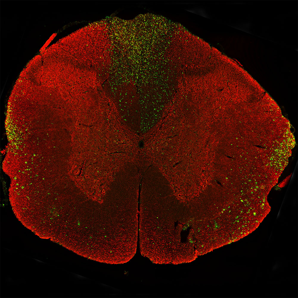
Above: Immunostaining of a coronal section of spinal cord of a rat given a midline C4 contusion injury three days previously. Sections were stained with RPCA-NF-L-ct in red and MCA-6H63 in green. RPCA-NF-L-ct binds an epitope in the C-terminal “tail” of NF-L which is destroyed or removed during degeneration. In contrast MCA-6H63 binds an epitope hidden in the core of the neurofilaments in uninjured neurons which are revealed on degeneration. MCA-6H63 stains prominent aggregates of material concentrated in the lateral funiculi and the dorsal columns but seen in lesser amounts throughout the section. These are degenerating and degenerated axonal processes which are generally negative for the the RPCA-NF-L-ct antibody. These two reagents are members of our novel panel of Degenotag™ reagents. Mouse select for larger image.
Publications making use of Degenotag™ reagents
1. Xin W wt al. Adolescent oligodendrogenesis and myelination restrict experience-dependent neuronal plasticity in adult visual cortex.
BioRχiv doi.org/10.1101/2023.09.29.560231 (2023).
2. Duncan DJ et al. Remyelination protects neurons from DLKmediated neurodegeneration.
BioRχiv doi.org/10.1101/2023.09.30.560267 (2023).