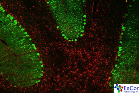
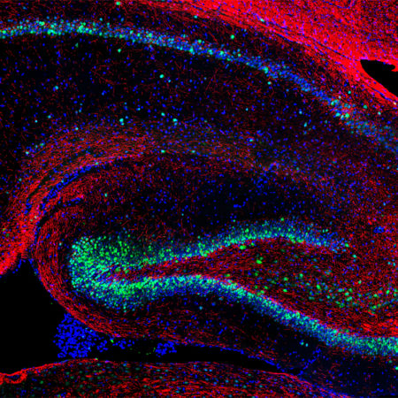
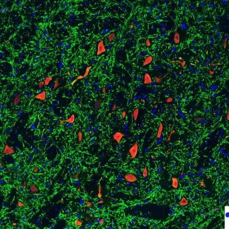
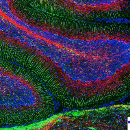
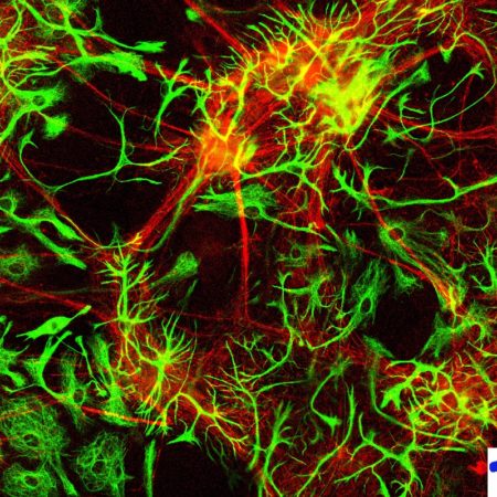
Adult rat cerebellum stained with CPCA-Calb, a chicken polyclonal antibody to the calcium binding protein calbindin in green and MCA-5C10, a mouse monoclonal antibody to glial fibrillary acidic protein (GFAP) in red. Calbindin is prominently expressed in the dendrites of Purkinje cells in the molecular layer.
Mouse select image above right for magnified view.
Image was taken on a Olympus FV1000 confocal microscope fitted with a programmable stage which allows a series of images to be taken and then merged into one large image. This produced a 14.1 megapixel image, will print on 24” X 36” paper at 128 DPI. To see a larger version of this image, as part of a slide show of our other images, go here.
Stained with CPCA-Calb, a chicken polyclonal antibody to the calcium binding protein calbindin in green.
Also MCA-5C10, a mouse monoclonal antibody to glial fibrillary acidic protein (GFAP) in red.
Image was taken on a Olympus FV1000 confocal microscope fitted with a programmable stage which allows a series of images to be taken and then merged into one large image. Cells were derived from E20 rat brain and were processed using our standard procedure outlined here.

