

EnCor Biotechnology
Chicken Polyclonal Antibody to Calbindin, Cat# CPCA-Calb
Description
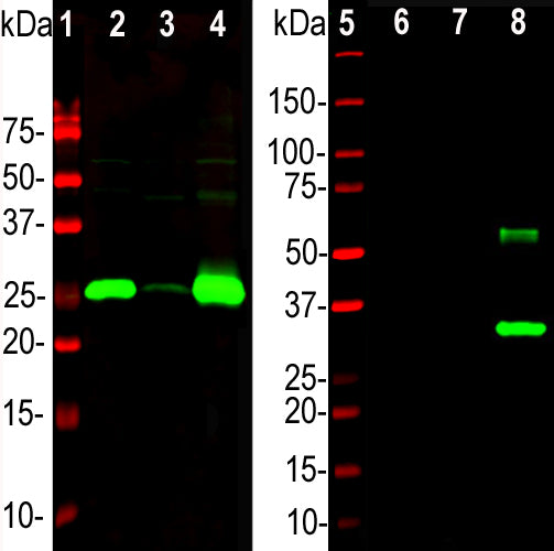
The CPCA-Calb antibody was made against full length recombinant human calbindin expressed in and purified from E. coli. We have shown that the antibody binds calbindin cleanly on western blots and in sections but fails to recognize the related calretinin and parvalbumin proteins. It is therefore ideally suited for identifying and subclassifying cortical GABAergic neurons. EnCor also manufactures mouse monoclonal antibodies to calbindin MCA-4H7 and MCA-5A9. We also supply a variety of other mouse, rabbit and chicken antibodies to parvalbumin and calretinin allowing double and triple labeling of appropriate cells and tissue specimens. Mouse select image above left for larger view.
Add a short description for this tabbed section
| Immunogen: | Full-length recombinant human protein expressed in and purified from E. coli. |
| HGNC Name: | CALB1 |
| UniProt: | P05937 |
| Molecular Weight: | 28kDa |
| Host: | Chicken |
| Species Cross-Reactivity: | Human, cow, rat, mouse |
| RRID: | AB_2572237 |
| Format: | Concentrated IgY preparation in PBS plus 0.02% NaN3 |
| Applications: | WB, IF/ICC, IHC |
| Recommended Dilutions: | WB: 1:5,000. IF/ICC 1:1,000-1:5,000. IHC: 1;5,000-1:10,000. |
| Storage: | Store at 4°C. Stable for 12 months from date of receipt. |
Calbindin, also known as calbindin 1 or calbindin-D28k is a member of the large superfamily of cytoplasmic EF hand containing Calcium binding proteins and is expressed in the brain, intestine, kidney and pancreas (1-3). It is particularly concentrated in the dendrites and perikarya of cerebellar Purkinje cells, but is also found in many GABAergic interneurons in the cerebral cortex. These GABAergic interneurons in most cases express only one of three Calcium binding proteins, namely calbindin or parvalbumin or calretinin. As a result these important and physiologically distinct inhibitory interneurons can be identified and subclassified based on their content of these three proteins (4-6).

Chromogenic immunostaining of a formalin fixed paraffin embedded rat cerebellum section with chicken pAb to calbindin, CPCA-Calb, dilution 1:5,000, detected in DAB (brown) following the ABC method with citrate buffer retrieval. Hematoxylin (blue) was used as the counterstain. Mouse select image for larger view.
1. Kretsinger RH, Nockolds CE. Carp Muscle Calcium-binding Protein: II. Structure determination and general description. J. Biol. Chem. 248:3313-26 (1973).
2. Andressen C, Bliimcke I, Celio MR. Calcium-binding proteins: selective markers of nerve cells. Cell Tissue Res. 271:181-208 (1993).
3. Schwaller B, Meyer M, Schiffmann S. ‘New’ functions for ‘old’ proteins: The role of the calcium binding proteins calbindin D-28k, calretinin and parvalbumin, in cerebellar physiology. Studies with knockout mice. The Cerebellum 1:241–58 (2002).
4. Celio MR. Calbindin D-28k and parvalbumin in the rat nervous system. Neurosci. 35:375-475 (1990).
5. Condé F, et al. Local circuit neurons immunoreactive for calretinin, calbindin D‐28k or parvalbumin in monkey prefronatal cortex: Distribution and morphology. J. Comp. Neurol. 341:95-116 (1994).
6. Hof PR, et al. Cellular distribution of the calcium-binding proteins parvalbumin, calbindin, and calretinin in the neocortex of mammals: phylogenetic and developmental patterns. J. Chem. Neuroanat. 16:77-116 (1999).
Add a short description for this tabbed section


