
EnCor Biotechnology
Chicken Polyclonal Antibody to CNP, Cat# CPCA-CNP
Description
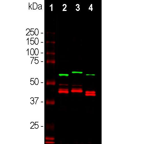
The CPCA-CNP antibody was made against the full length recombinant form of human CNP expressed in and purified from E. Coli, and the antibody can be used to identify myelinating cells in cell culture and in sections and to trace axonal projections in sectioned material. We document that the antibody works well not only for western blotting, IF and ICC but also on formalin fixed paraffin embedded sections of human and rodent tissues, select the "Additional Data" for this data. The same recombinant protein was used to generate polyclonal rabbit and goat antibodies to CNP RPCA-CNP and GPCA-CNP and also a mouse monoclonal MCA-1H10. These antibodies are excellent markers of myelin and myelinating cells and recognize CNP cleanly on western blots.
- Cell Structure Marker
- Cell Type Marker
- Chicken Polyclonal Antibodies
- Cytoplasmic Enzyme
- Immunohistochemistry Verified
Add a short description for this tabbed section
| Immunogen: | Full length recombinant human CNP protein |
| HGNC Name: | CNP |
| UniProt: | P09543 |
| Molecular Weight: | 46kDa, 48kDa |
| Host: | Chicken |
| Species Cross-Reactivity: | Human, rat, mouse, cow, pig, horse |
| RRID: | AB_2572249 |
| Format: | Concentrated IgY preparation plus 0.02% NaN3 |
| Applications: | WB, ICC/IF, IHC |
| Recommended Dilutions: | WB: 1:5,000-10,000. IF/IHC: 1:2,000-4,000. |
| Storage: | Store at 4°C. Stable for 12 months from date of receipt. |
The 2′,3′-cyclic nucleotide 3′-phosphodiesterase (CNP), is an enzyme which catalyzes the hydrolysis of 2′, 3′-cyclic nucleotides to 2′-nucleotides. These cyclic nucleotides are structurally different from the better known and studied 3′-5′-cyclic nucleotides of which the best known example is cyclic AMP. CNP has two isoforms, CNPase 1 (46kDa) and CNPase 2 (48kDa), which are encoded separately by different promoters of the same gene (1). These enzymes are present in very high levels in brain and peripheral nerve, makes up 4% of total CNS myelin protein. They are found almost exclusively in oligodendrocytes and Schwann cells, appearing early in oligodendrocyte development, earlier than most other myelin specific proteins (2). Antibodies to CNP have been very useful as a marker for these particular cell types. CNP is thought to play a critical role in the events leading up to myelination, for the oligodendrocytes overexpressing CNP appear to mature earlier in development, resulting in earlier maximum gene expression for myelin basic proteins (3). It has been reported that CNP is also associated with microtubules in brain tissue and may promote microtubule assembly. CNP can link tubulin to cellular membranes, and may regulate cytoplasmic microtubule distribution (4). In various diseases, neurological mutants, and in experimental conditions in which myelin is reduced, CNP levels may also be severely reduced. Decreased brain levels of CNP have also been reported in Down syndrome and Alzheimer’s disease (5).

Chromogenic Immunostaining of a formalin fixed paraffin embedded human cerebral cortex tissue section with chicken pAb to CNP, CPCA-CNP, dilution 1:4,000, detected in DAB (brown) following the ABC method with citrate buffer retrieval at pH=6.0. Hematoxylin (blue) was used as the counterstain. The CNP antibody stains myelin and oligodendrocytes, cells that produce the myelin sheath around axons. Mouse select image for larger view.
1. Monoh K, Kurihara T, Sakimura K, Takahashi Y. Structure of mouse 2′,3′-cyclic-nucleotide 3′-phosphodiesterase gene. BBRC 165:1213-20 (1989).
2. Kasama-Yoshida H, et al. A comparative study of 2′,3′-cyclic-nucleotide 3′-phosphodiesterase in vertebrates: cDNA cloning and amino acid sequences for chicken and bullfrog enzymes. J. Neurochem. 69:1335–42 (1997).
3. Gravel M, et al. Overexpression of 2′,3′-cyclic nucleotide 3′-phosphodiesterase in transgenic mice alters oligodendrocyte development and produces aberrant myelination. Mol. Cell. Neurosci. 6:453-66 (1996).
4. Bifulco M, Laezza C, Stingo S, Wolff J. 2′,3′-Cyclic nucleotide 3′-phosphodiesterase: a membrane-bound, microtubule-associated protein and membrane anchor for tubulin. PNAS 99:1807–11 (2001).
5. Vlkolinský R, Cairns N, Fountoulakis M, Lubec G. Decreased brain levels of 2′,3′-cyclic nucleotide-3′-phosphodiesterase in Down syndrome and Alzheimer’s disease. Neurobiol. Aging 22:547-53 (2001).
Add a short description for this tabbed section


