
EnCor Biotechnology
Mouse Monoclonal Antibody to Glial Fibrillary Acidic Protein-Human (GFAP), Cat# MCA-2A5
Description
The MCA-2A5 antibody was raised against GFAP purified from pig spinal cord. It works well on human tissues and is particularly useful as a capture reagent in ELISA. As shown at the left it also works well for immunohistochemistry on paraffin sections of human material. The MCA-2A5 epitope is somewhat divergent between human and rodents, so, while the antibody does work, it is not optimal for rodent IF/ICC and IHC studies, for those species we recommend MCA-5C10. We also used recombinant human GFAP to generate rabbit, RPCA-GFAP, chicken CPCA-GFAP and goat, GPCA-GFAP polyclonal antibodies to GFAP. These all work well for WB, IF/ICC and IHC on material from human, rodent and other mammalian species.
- Cell Structure Marker
- Cell Type Marker
- Cytoskeletal Marker
- Developmental Marker
- Epitope Mapped Antibodies
- Immunohistochemistry Verified
- Mouse Monoclonal Antibodies
Add a short description for this tabbed section
| Immunogen: | GFAP isolated biochemically from pig spinal cord |
| HGNC Name: | GFAP |
| UniProt: | F1RR02 |
| Molecular Weight: | 50kDa |
| Host: | Mouse |
| Isotype: | IgG1 |
| Species Cross-Reactivity: | Human, cow, pig, weak on rat and mouse |
| RRID: | AB_2732880 |
| Format: | Protein G affinity purified antibody at 1mg/mL in 50% PBS, 50% glycerol plus 5mM NaN3 |
| Applications: | WB, IF/ICC, IHC |
| Recommended Dilutions: | WB: 1:2,000. IF/ICC: 1:500. IHC: 1:2,000. |
| Storage: | Store at 4°C for short term, for longer term store at -20°C. Stable for 12 months from date of receipt. |
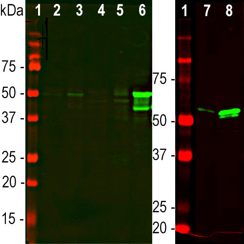
Glial Fibrillary Acidic Protein (GFAP) is strongly and specifically expressed in astrocytes, Bergmann glia and certain other glia in the central nervous system, in satellite cells in peripheral ganglia, and in non-myelinating Schwann cells in peripheral nerves (1-3). GFAP expression is also seen in developing neural stem cells and GFAP levels may greatly increase in regions of CNS injury or disease (4), and point mutations in the GFAP gene are causative of Alexander's disease (5). Antibodies to GFAP such as MCA-2A5 are useful for visualizing glia and monitoring developmental, disease and damage related CNS alterations. This antibody has been shown to work well on western blots, IF, ICC and IHC of human. pig and cow samples. Some interest has recently been focused on GFAP as a protein released into blood and CSF following traumatic brain injury, stroke and other CNS compromises (6). Measurement of the levels of blood or CSF GFAP may give information about patient presention, progress, response to therapy or outcome. MCA-2A5 has been widely used as a capture reagent in ELISA and other antibody based assays detecting human GFAP in human blood and CSF samples. MCA-2A5 also works well on paraffin sections of human brain, see data under the "additional info" tag.
This antibody was made against purified pig spinal cord GFAP and works well on human tissues but is less efficient on rodent material. The epitope for this antibody has been mapped to the N-terminal region of the α-helical coiled-coil "rod" region of human GFAP isotype I, and further studies show that the human peptide EIRFLRKIHEEEVRE (196-210) strongly inhibits binding of the antibody to human GFAP. This peptide is conserved between pig and human GFAP, but that of rat and mouse are both slightly divergent, having a glutamine instead of the arginine (see below). This small divergence is likely the reason for the lower efficiency of this antibody on rodent material.
Human EIRFLRKIHEEEVRE (196-210)
Rat EIQFLRKIHEEEVRE (194-208)
Mouse EIQFLRKIYEEEVRE (193-207)

Immunofluorescent analysis of an adult rat cerebellum section stained with mouse mAb to GFAP, MCA-2A5, dilution 1:500, in red, and costained with chicken pAb to parvalbumin, CPCA-Pvalb, dilution 1:2,000, in green. The blue is DAPI staining of nuclear DNA. Following transcardial perfusion of rat with 4% paraformaldehyde, brain was post fixed for 24 hours, and free-floating 45μM sections were stained with above antibodies. The GFAP antibody stains the processes of Bergmann glia and astrocytes, though is not optimal for this purpose, likely because the epitope is slightly variant in rodent compared to the human and the pig sequence. Mouse select image for larger view.
1. Bignami A, Eng LF, Dahl D, Uyeda CT. Localization of the glial fibrillary acidic protein in astrocytes by immunofluorescence. Brain Res. 43:429-35 1972.
2. Yen SH, Fields KL. Antibodies to neurofilament, glial filament, and fibroblast intermediate filament proteins bind to different cell types of the nervous system. J Cell Biol. 88:115-26 1981.
3. Shaw G, Osborn M, Weber K. An immunofluorescence microscopical study of the neurofilament triplet proteins, vimentin and glial fibrillary acidic protein within the adult rat brain. Eur J Cell Biol. 26:68-82 1981.
4. Fitch MT, Silver J. CNS injury, glial scars, and inflammation: Inhibitory extracellular matrices and regeneration failure. Exp Neurol. 209:294-301 2008.
5. Brenner M, et al. Mutations in GFAP, encoding glial fibrillary acidic protein, are associated with Alexander disease. Nat Genet 27:117-20 2001.
6. Schiff L, Hadker N, Weiser S, Rausch C. A literature review of the feasibility of glial fibrillary acidic protein as a biomarker for stroke and traumatic brain injury. Mol. Diagn. Ther. 16:79-92 (2012).
Add a short description for this tabbed section


,%20Cat%23%20MCA-2A5)