

EnCor Biotechnology
Rabbit Polyclonal Antibody to Adenylate Cyclase III (ACIII, AC111, AC3), Cat# RPCA-ACIII
Description
The RPCA-ACIII antibody was made against the extreme C-terminal peptide of rat ACIII, PAAFPNGSSVTLPHQVVDNP, amino acids 1125-1144 of the GenBank entry NP_570135.2. A cysteine residue was added to the N-terminus to allow coupling to MBS-activated keyhole limpet hemocyanin. The antibody works on mouse cells which express the same peptide and also on human cells, presumably because the corresponding peptide in the human ACIII sequence is the closely related peptide LATFPNGPSVTLPHQVVDNS. The antibody binds full length transfected human ACIII on western blots and works well to identify neuronal cilia on human and rodent cells. The antibody has become widely very used in peer reviewed publications, see here. We have also generated a mouse monoclonal and a chicken polyclonal antibody to the same ACIII peptide, MCA-1A12 and CPCA-ACIII.
- Cell Structure Marker
- Cell Type Marker
- Cytoskeletal Marker
- Epitope Mapped Antibodies
- Immunohistochemistry Verified
- Our Most Widely Used Reagents
- Rabbit Polyclonal Antibodies
Add a short description for this tabbed section
| Immunogen: | C-terminal peptide of rat ACIII, PAAFPNGSSVTLPHQVVDNP with a Cys added to the N-terminus to allow coupling to KLH. |
| HGNC Name: | ADCY3 |
| UniProt: | P21932 |
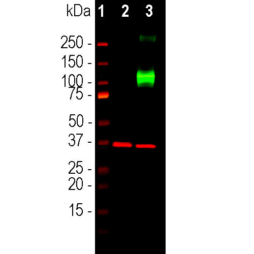
| Molecular Weight: | 130-200kDa by SDS-PAGE |
| Host: | Rabbit |
| Species Cross-Reactivity: | Human, rat, mouse |
| RRID: | AB_2572219 |
| Format: | Immunogen affinity purified antibody at 1mg/mL in 50% PBS, 50% glycerol plus 5mM NaN3 |
| Applications: | WB, IF/ICC |
| Recommended Dilutions: | WB: 1:1,000. IF/ICC: 1:2,000-5,000. |
| Storage: | Store at 4°C for short term, for longer term store at -20°C. Stable for 12 months from date of receipt. |
G-protein coupled receptors are a large and variable family of membrane proteins. On binding their specific ligand they activate specific members of the family of trimeric G-proteins which in turn activate other signaling enzymes. Adenylate cyclases are one of these and are activated by the GTP bound GαS subunits of trimeric G-proteins. Adenylate cyclases are responsible for the production of the important “second messenger” signaling molecule cyclic-AMP which in turn activates the cAMP dependent protein kinase. This kinase phosphorylates numerous substrate molecules on serine or threonine residues and so alters their activity. There are several different adenylate cyclase genes and protein products with each have distinctly different distribution patterns in cells and tissues. The type III adenylate cyclase enzyme is specifically localized in the membranes surrounding neuronal cilia, and is activated by specific G-protein coupled receptors also located in cilia (1-5). Neuronal cilia express a variety of other receptors types and mediators of other signaling pathways and appear to function as a unique and complex neuronal sensory structure (1-5). For examples, the somatostatin 3 receptor, neuropeptide Y 2 receptor and melanin concentrating hormone receptor 1 are localized in neuronal cilia and the sonic hedgehog and Wnt signaling pathway act on neurons primarily through neuronal cilia (6).
1. Fuchs JL, Schwark HD. Neuronal primary cilia: a review. Cell Biol. Int. 28:111-8 (2004).
2. Louvi A and Grove EA. Cilia in the CNS: the quiet organelle claims center stage. Neuron 69:1046-60 (2011).
3. Singla V, Reiter JF. The primary cilium as the cell's antenna: signaling at a sensory organelle. Science 313:629-33 (2006).
4. Green JA, Mykytyn K. Neuronal Primary Cilia: An Underappreciated Signaling and Sensory Organelle in the Brain. Neuropsychopharm. 39:244–5 (2014).
5. May-Simera HL, Kelley MW. Cilia, Wnt signaling, and the cytoskeleton. Cilia 2;1:7 (2012).
6. Guemez-Gamboa A, et al. Primary cilia in the developing and mature brain. Neuron 82:511-21 (2014).
7. Guadiana SM, et al. Arborization of Dendrites by developing neocortical neurons is dependent on primary cilia and Type 3 adenylyl cyclase. J. Neurosci. 33:2626-38 (2013).
This antibody has been cited in peer reviewed literature, see the CiteAb link here.
Add a short description for this tabbed section


,%20Cat%23%20RPCA-ACIII)