


EnCor Biotechnology
Chicken Polyclonal Antibody to Neurofilament NF-H (Nfh, NEFH), Cat# CPCA-NF-H
Description
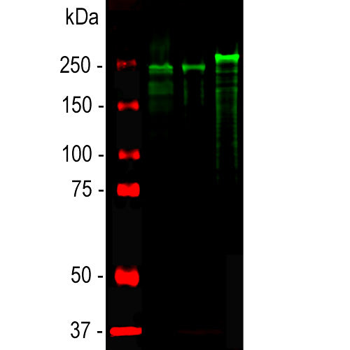
The CPCA-NF-H antibody was raised against biochemically isolated NF-H purified from bovine spinal cord (9). This preparation is dominated by axonal forms of NF-H which are heavily phosphorylated on the multiply repeated NF-H KSP type sequences, and this antibody reacts very strongly with these phosphorylated repeats. Reactivity with non-phosphorylated KSP sequences is orders of magnitude weaker, similar to other characterized antibodies to NF-H (5). In most species there is some cross-reactivity with the phosphorylated KSP sequences found in the related neurofilament subunit NF-M which are similar but not identical to those of NF-H. The antibody recognizes phosphorylated NF-H strongly in all mammals tested to date and also in chicken and has an unusually high titer of 1:20,000 or higher. We document that the antibody works well not only for western blotting, IF and ICC but also on formalin fixed paraffin embedded sections of human and rodent tissues, select the "Additional Data" for this data. The antibody is widely used and sold through many vendors, see for example the results of Google Scholar search for CPCA-NF-H. We also market mouse monoclonal antibodies MCA-NAP4, MCA-9B12 and MCA-AH1 and rabbit polyclonal and goat polyclonal antibodies RPCA-NF-H and GPCA-NF-H all of which have similar specificity to CPCA-NF-H.
- Cell Structure Marker
- Cell Type Marker
- Chicken Polyclonal Antibodies
- Cytoskeletal Marker
- Developmental Marker
- Immunohistochemistry Verified
Add a short description for this tabbed section
| Immunogen: | Native NF-H purified from bovine spinal cord. |
| HGNC Name: | NEFH |
| UniProt: | P12036 |
| Molecular Weight: | 200-220kDa by SDS-PAGE |
| Host: | Chicken |
| Species Cross-Reactivity: | Human, rat, mouse, cow, pig, dog, horse |
| RRID: | AB_2149761 |
| Format: | Concentrated IgY preparation in PBS plus 0.02% NaN3 |
| Applications: | WB, IF/ICC, IHC, ELISA |
| Recommended Dilutions: | WB: 1:20,000-1:50,000. IF/ICC, and IHC: 1:5,000-1:10,000. |
| Storage: | Store at 4°C. Stable for 12 months from date of receipt. |
Neurofilaments are the 10nm or intermediate filament proteins found specifically in neurons, and are composed predominantly of three major proteins called NF-L, NF-M and NF-H, though other proteins may also be present. NF-H is the neurofilament high or heavy molecular weight polypeptide and runs on SDS-PAGE gels at 200-220 kDa, with some variability across species boundaries. The protein is in reality much smaller in molecular size, about 110kDa (1,2). The unusual SDS-PAGE mobility is due partly to a very high content of charged amino acids, particularly glutamic acid rich regions, and the non-phosphorylated form runs on SDS-PAGE at about 160kDa. The predominant type of NF-H is the axonal form which is heavily serine phosphorylated on 40 or more tandemly repeated lysine-serine-proline (KSP) containing peptides (3-5). The phosphorylation of these peptides results in considerable further retardation on SDS-PAGE gels, so the heavily phosphorylated axonal form runs at 200-220kDa with some species variability. Antibodies to NF-H are useful for identifying axonal processes in tissue sections and in culture. NF-H antibodies can also be useful in visualizing neurofilament accumulations seen in many neurological disorders, such as Amyotrophic Lateral Sclerosis (also known as Lou Gehrig’s disease), Alzheimer’s disease and following traumatic injury. The phosphorylated axonal form of NF-H, usually referred to as pNF-H, can be detected in blood and CSF following a variety of damage and disease states resulting in axonal compromise, and antibodies such as this can be used to used to quantify such ongoing axonal loss (e.g. 6-8).

Chromogenic immunostaining of a formalin fixed paraffin embedded rat cerebellum section with chicken pAb to NF-H, CPCA-NF-H, dilution 1:10,000, detected with DAB (brown) following the ABC method with citrate buffer retrieval.. Hematoxylin (blue) was used as the counterstain. The NF-H antibody labels axonal profiles of basket cells surrounding Purkinje neurons and processes from other types of neuron. Mouse select image for larger view.
This antibody has become widely used as sold by EnCor and through our numerous OEM partners, and in-formation on this can be viewed using Google scholar by searching for "CPCA-NF-H” or by selecting here. Here is a CiteAb link to peer reviewed publications which use this antibody obtained directly from EnCor, here.
A considerable literature now shows that detection of pNF-H in plasma, serum and CSF is a useful surrogate marker of axonal loss resulting from CNS damage disease (4). This antibody in the antigen affinity purified form has been used as a very effective ELISA capture reagent and is a component of our ELISA-pNF-H-v1 kit.
1. Perrot R, et al. Review of the Multiple Aspects of Neurofilament Functions, and their Possible Contribution to Neurodegeneration. Mol. Neurobiol. 38:27-65 (2008).
2. Lépinoux-Chambaud C. Eyer J. Review on intermediate filaments of the nervous system and their pathological alterations. Histochem. Cell Biol. 140:13-22 (2013).
3. Sternberger LA, Sternberger NH. Monoclonal antibodies distinguish phosphorylated and nonphosphorylated forms of neurofilaments in situ. PNAS
80:6126-30 (1983).
4. Julien JP, Mushynski WE. Multiple phosphorylation sites in mammalian neurofilament polypeptides. J. Biol. Chem. 257:10467-70 (1982).
5. Lee VM, et al. Identification of the major multiphosphorylation site in mammalian neurofilaments. PNAS 85:1998-2002 (1988).
6. Shaw G, et al. Hyperphosphorylated neurofilament NF-H is a serum biomarker of axonal injury. Biochem. Biophys. Res. Commun. 336:1268-77 (2005).
7. Boylan et al, Immunoreactivity of the phosphorylated axonal neurofilament H subunit (pNF-H) in blood of ALS model rodents and ALS patients: evaluation of blood pNF-H as a potential ALS biomarker. J. Neurochem. 111:1182-91 (2009).
8. Shaw G. The Use and Potential of pNF-H as a General Blood Biomarker of Axonal Loss: An Immediate Application for CNS Injury. In: Kobeissy FH, editor. Brain Neurotrauma: Molecular, Neuropsychological, and Rehabilitation Aspects. CRC Press/Taylor & Francis; 2015. Chapter 21 .
9. Delacourte A, et al. Study of the 10-nm-filament fraction isolated during the standard microtubule preparation. Biochem. J. 191:543-6 (1980).
Add a short description for this tabbed section


,%20Cat%23%20CPCA-NF-H)