

EnCor Biotechnology
Chicken Polyclonal Antibody to Neurofilament NF-L (Nfl, NEFL) DegenoTag™ Peptide, Cat# CPCA-NF-L-Degen
Description
The CPCA-NF-L-Degen was raised against a proprietary recombinant immunogen containing amino acids 311-375 of the human NF-L sequence and is one of our DegenoTag™" reagents which specifically binds to degenerated forms of NF-L. Full details of these findings are described in a peer-reviewed publication in Brain Communications. The antibody works well on western blots of a variety of species but binds only degenerated processes in sectioned material. We recommend using the antibody at 1:10,000 dilution for the specific visualization of degenerating and degenerated processes by IF/ICC. It also works well on paraffin embedded histological sections of rodent CNS tissues, including transgenic mouse models. This and other DegenoTag™ reagents can be used to identify degenerating and degenerated processes and also to monitor NF-L degradation in a variety of contexts. EnCor generated another polyclonal DegenoTag™ reagent made in rabbit RPCA-NF-L-Degen. We also generated several monoclonal antibodies specific for degenerated forms of NF-L, MCA-6H63, MCA-1D44 and MCA-1B11.
- Cell Structure Marker
- Cell Type Marker
- Chicken Polyclonal Antibodies
- Developmental Marker
- Epitope Mapped Antibodies
- Immunohistochemistry Verified
- New Antibodies
Add a short description for this tabbed section
| Immunogen: | Degenotag™ NF-L peptide |
| HGNC Name: | NEFL |
| UniProt: | P07196 |
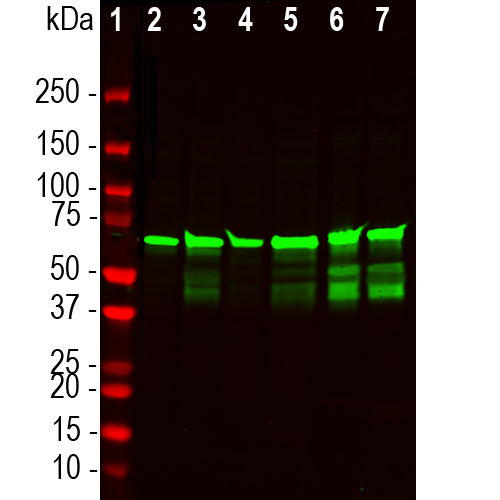
| Molecular Weight: | 68-70kDa |
| Host: | Chicken |
| Species Cross-Reactivity: | Human, rat, mouse, pig, cow |
| RRID: | AB_2923500 |
| Format: | Immunogen affinity purified antibody at 1mg/mL in 50% PBS, 50% glycerol plus 5mM NaN3 |
| Applications: | WB, IF/ICC, IHC |
| Recommended Dilutions: | WB: 1:10,000-1:20,000. IF/ICC: 1:5,000-1:10,000. IHC: 1:10,000-1:20,000. |
| Storage: | Store at 4°C for short term, for longer term store at -20°C. Stable for 12 months from date of receipt. |
Neurofilaments are major components of neurons and their axons (1-5). We have recently developed a series of novel reagents which we call DegenoTag™ products. These are antibodies which recognize epitopes in a small segment of the neurofilament NF-L subunit which are normally not accessible to antibodies but which became available on degeneration. We propose that these epitopes are made accessible as a result of degeneration induced proteolysis, and in agreement with this hypothesis we could make previously negative control tissues become strongly DegenoTag™ antibody positive by treatment with proteases. In addition fresh CNS tissues did not stain with DegenoTag™ reagents except for a tiny minority of apparently spontaneously degenerating processes. In stark contrast tissues left to sit at room temperature for 4 hours were strongly reactive with DegenoTag™ reagents. We also discovered that our antibodies to the C-terminal of NF-L, such as our rabbit polyclonal RPCA-NF-L-ct and mouse monoclonal MCA-DA2. Our reagents can therefore be used to positively identify both healthy and degenerated processes

Chromogenic immunohistochemistry on a formalin fixed paraffin embedded spinal cord section of an ALS mouse model from the lab of Dr. David Borchelt (6). These heterozygous G85R-SOD1-YFP mice do not develop ALS unless they are primed by injection of mutant SOD1 aggregates, so this is a model of prion induction of pathogenesis. This mouse was injected with mutant SOD1 in the hind limb and degenerated processes were seen in the sciatic nerve, in fibers in the spinal cord and as shown here in the brain stem. Staining was performed with chicken pAb to degenerated neurofilament NF-L, CPCA-NF-L-Degen, dilution 1:10,000, detected with DAB (brown) using the ABC method and reagents with no antigen retrieval.Hematoxylin (blue) was used as the counterstain. Hematoxylin (blue) was used as the counterstain. Mouse select image for larger view.
1. Hoffman et al. Neurofilament gene expression:a major determinant of axonal caliber. PNAS 84:3472-6 (1987).
2. Perrot R, et al. Review of the Multiple Aspects of Neurofilament Functions, and their Possible Contribution to Neurodegeneration. Mol. Neurobiol. 38:27-65 (2008).
3. Lépinoux-Chambaud C. Eyer J. Review on intermediate filaments of the nervous system and their pathological alterations. Histochem. Cell Biol. 140:13-22 (2013).
4. Liu Q. et al. Neurofilamentopathy in Neurodegenerative Diseases. Open Neurol. J. 5:58–62 (2011).
5. Bacioglu M, et al. Neurofilament light chain in blood and CSF as marker of disease progression in mouse models and in neurodegenerative diseases. Neuron 91:56-66 (2016).
6. Shaw G, et al. Uman Type NF-L Antibodies Are Effective Reagents for the Imaging of Neurodegeneration. BioRχiv DOI 10.1101/2022.08.27.504533 (2022).
7. Norgren N, et al. Monoclonal antibodies selective for low molecular weight neurofilaments. Hybrid Hybridomics 21:53-59 (2002).
Add a short description for this tabbed section


%20DegenoTag%26trade;%20Peptide,%20Cat%23%20CPCA-NF-L-Degen)