

EnCor Biotechnology
Chicken Polyclonal Antibody to Peripherin, Cat# CPCA-Peri
Description
The CPCA-Peri antibody was made against full length recombinant rat peripherin expressed in and purified from E. coli. It recognizes peripherin from a variety of species on western blots and we document that it works well for, IF and ICC and also on formalin fixed paraffin embedded sections, select the "Additional Data" for this data. We also manufacture a mouse monoclonal antibody to peripherin which has been used in studies of peripherin expression for many years in many peer reviewed publications, MCA-8G2 which recognizes both the human and the rodent protein. We also made a rabbit polyclonal antibody to peripherin, RPCA-Peri.. Finally we have made a mouse monoclonal antibody to peripherin which recognizes rodent but not human peripherin, MCA-7C5.
- Cell Type Marker
- Chicken Polyclonal Antibodies
- Cytoskeletal Marker
- Developmental Marker
- Immunohistochemistry Verified
Add a short description for this tabbed section
| Immunogen: | Recombinant rat peripherin expressed in and purified from E. coli |
| HGNC Name: | PRPH |
| UniProt: | P21807 |
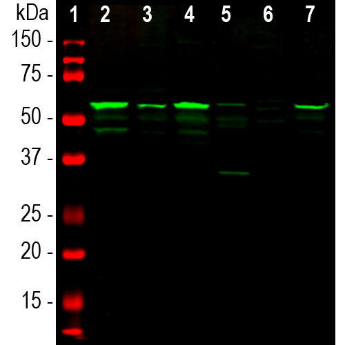
| Molecular Weight: | 57kDa |
| Host: | Chicken |
| Species Cross-Reactivity: | Human, Rat, Mouse, Pig, Cow |
| RRID: | AB_2284443 |
| Format: | Concentrated IgY preparation in PBS plus 0.02% NaN3 |
| Applications: | WB, IF/ICC, IHC |
| Recommended Dilutions: | WB: 1:20,000. IF/ICC: 1:2,000-1:5,000. IHC: 1:20,000. |
| Storage: | Stable at 4°C for at least one year. Mix 1:1 with 100% glycerol and store at -20°C for longer term storage |
Peripherin is a ~57kDa intermediate filament subunit found initially in sensory neurons of the peripheral nervous systems, which gives the protein its name (1). Subsequently, peripherin was found in some sensory and other neurons of the central nervous system and also as a major cytoskeletal protein in rat pheochromacytoma PC12 cells (2,3). Peripherin is expressed in certain neuroendocrine cells and in the insulin producing cells of the pancreas, and autoantibodies to peripherin may be seen in diabetes mellitus (4). It is also prominently expressed in certain tumors and in the ballooned axons typical of amyotrophic lateral sclerosis (5,6). Different classes of neuron and nerve fiber may be identified based on their expression of peripherin, NF-L and other markers (e.g. 7). There are multiple transcripts from the single peripherin gene, some of which encode proteins which produce proteins unable to form intermediate filaments and which induce the formation of pathological inclusions (8,9). Note that the intermediate filament subunit peripherin (HGNC PRPH) is completely unrelated to peripherin/RDS (HGNC PRPH2), a retinal protein belonging to the tetraspanin family.

Chromogenic immunostaining of a formalin fixed paraffin embedded rat brain stem section with chicken pAb to peripherin, CPCA-Peri, dilution 1:20,000, detected with DAB (brown) following the ABC method with citrate buffer retrieval. Hematoxylin (blue) was used as the counterstain. In the central nervous system, peripherin antibody primarily labels neurons which have projections into the peripheral nervous system. Mouse select image for larger view.

Chromogenic immunostaining of a formalin fixed paraffin embedded rat brain stem section with chicken pAb to peripherin, CPCA-Peri, dilution 1:20,000, detected with DAB (brown) following the ABC method with citrate buffer retrieval. Hematoxylin (blue) was used as the counterstain. In the central nervous system, peripherin antibody labels axonal bundles which project into the peripheral nervous system. Mouse select image for larger view.
1. Portier MM, de Néchaud B, Gros F. Peripherin, a new member of the intermediate filament protein family. Dev. Neurosci. 6:335-44 (1984).
2. Troy CM, Brown K, Greene LA, Shelanski ML. Ontogeny of the neuronal intermediate filament protein, peripherin, in the mouse embryo. Neuroscience 36:217-37 (1990).
3. Aletta JM, et al. Relationship between the nerve growth factor-regulated clone 73 gene product and the 58-kilodalton neuronal intermediate filament protein (peripherin). J. Neurochem 51:1317-20 (1988).
4. Puertas MC, et al. Peripherin Is a Relevant Neuroendocrine Autoantigen Recognized by Islet-Infiltrating B Lymphocytes. J. Immunol. 178:6533-9 (2007).
5. Migheli A, et al. Peripherin immunoreactive structures in amyotrophic lateral sclerosis. Lab. Invest. 68:185-91 (1993).
6. He CZ, Hays AP. Expression of peripherin in ubiquinated inclusions of amyotrophic lateral sclerosis. J. Neurol. Sci. 217:47-54 (2004).
7. Goldstein ME, House SB, Gainer H. NF‐L and peripherin immunoreactivities define distinct classes of rat sensory ganglion cells. J. Neurosci. Res. 30:92-104 (1991).
8. Landon F, et al. Multiple mRNAs encode peripherin, a neuronal intermediate filament protein. EMBO J. 8:1719-26 (1989).
9. Robertson J, et al. A neurotoxic peripherin splice variant in a mouse model of ALS. J. Cell Biol. 60:939-49 (2003).
Add a short description for this tabbed section


