


EnCor Biotechnology
Chicken Polyclonal Antibody to Vimentin, Cat# CPCA-Vim
Description
The CPCA-Vim antibody can be used to study stem cells and generally to reveal the intermediate filament cytoskeleton. The antibody works well for IF and ICC, but is not recommended for IHC. The immunogen used to generate this antibody was full length recombinant human vimentin, PROT-r-Vim, expressed in and purified from E. coli. The antibody works well on all mammals tested to date, and it was generated in chicken by standard procedures and immunoglobulin was extracted from egg yolk. The resulting polyclonal antibody belongs to the IgY subclass, the chicken homolog of mammalian IgG and can be used in the same way, with the caveat that this type of antibody does not bind either Protein A or Protein G. Suitable second antibody reagents can be obtained from many vendors including ThermoFisher and Sigma-Aldrich. The same vimentin immunogen was used to produce two high quality epitope mapped monoclonal antibodies to vimentin MCA-2A52 and MCA-2D1, and also a rabbit polyclonal antibody RPCA-VIM.
- Cell Structure Marker
- Cell Type Marker
- Chicken Polyclonal Antibodies
- Cytoskeletal Marker
- Developmental Marker
- Immunohistochemistry Verified
Add a short description for this tabbed section
| Immunogen: | Full length recombinant human vimentin protein , PROT-r-Vim, expressed in and purified from E. coli. expressed in and purified from E. coli. |
| HGNC Name: | VIM |
| UniProt: | P08670 |
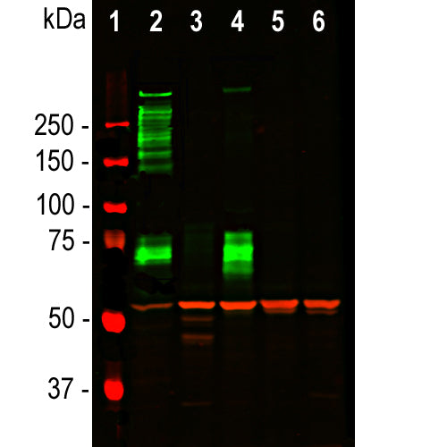
| Molecular Weight: | 50kDa |
| Host: | Chicken |
| Species Cross-Reactivity: | Human, rat, mouse, cow, pig, horse, chicken |
| RRID: | AB_2216401 |
| Format: | Concentrated IgY preparation in PBS plus 0.02% NaN3 |
| Applications: | WB, IF/ICC |
| Recommended Dilutions: | WB: 1:5,000. IF/ICC: 1:10,000. IHC not recommended. |
| Storage: | Store at 4°C. Stable for 12 months from date of receipt. |
Vimentin is a protein subunit of the intermediate or 10nm filaments found in the cytoplasm of many cell types (1). Intermediate filaments are relatively stable fibrous components of cells which appear to have primarily a mechanical function. Many cell lines such as HEK293, HeLa, 3T3 and Cos cells contain prominent vimentin networks (1). Vimentin is a major protein of eye lens and cornea, and found generally in mesenchymal tissues in adult mammals. In the CNS it is found in endothelia and developing neurons, developing and some mature astrocytes, microglia, mature Bergmann glia and ependyma (2,3). Mutations in the vimentin gene may cause cataracts (4,5), and elevated levels of vimentin in blood samples are associated with onset of cancer (6,7). Vimentin levels increase in a variety of cell types as they become cancerous, suggesting that increase in expression of this protein is a useful diagnostic marker of the epithelial-mesenchymal transition, an important step in the metastasis of carcinoma cells (8).

Chromogenic Immunostaining of a 4% PFA fixed paraffin embedded rat kidney section with chicken pAb to vimentin, CPCA-Vim, dilution 1:5,000, detected with DAB (brown) following the ABC method with citrate buffer retrieval at pH=6.0. Hematoxylin (blue) was used as the counterstain. The RPCA-Vim antibody specifically labels the interstitium in glomerular podocytes. On brain sections, only endothelium stained positively. This antibody performs well in testing with 4% PFA but is not recommended for standard NBF fixed tissues. Mouse select image for larger view.

Neuromics hN2 are human neuronal lineage cells which originate from ArunA Biomedical, and were derived from the human embryonic cell line WA09. We obtained the hN2 Human Neurons from Neuromics, which arrived frozen and on dry ice. We fixed and stained them with our chicken polyclonal antibody to vimentin CPCA-Vim, in red. Islands of hN2 cells form after 4 days in culture forming beautiful flower like structures. Vimentin is a well established marker of early differentiating neuronal lineage cells, though it is found in many other kinds of cells. Merged from several images taken with a 10X objective lens, this image is also available as a high resolution poster. Blue staining is nuclear DNA. Mouse select image for larger view.
1. Franke WW, et al. Different intermediate-sized filaments distinguished by immunofluorescence microscopy. PNAS 75:5034–8 (1978).
2. Dahl D, et al. Vimentin, the 57 000 molecular weight protein of fibroblast filaments, is the major cytoskeletal component in immature glia. Eur. J. Cell Biol. 24:191-6 (1981).
3. Shaw, G. et al. An immunofluorescence microscopical study of the neurofilament triplet proteins, vimentin and glial fibrillary acidic protein within the adult rat brain. Eur. J. Cell Biol. 26:68-72 (1981).
4. Muller M, et al. Dominant cataract formation in association with a vimentin assembly disrupting mutation. Hum. Molec. Genet. 18:1052-7 (2009).
5. Zhai Y, et al. Targeted exome sequencing of congenital cataracts related genes: broadening the mutation spectrum and genotype-phenotype correlations in 27 Chinese Han families. Sci. Rep. 7:1219 (2017).
6. Satelli A, Li S. Vimentin in cancer and its potential as a molecular target for cancer therapy. Cell Mol. Life Sci. 68:3033-46 (2011).
7. Wong KF, Luk JM. Discovery of lamin B1 and vimentin as circulating biomarkers for early hepatocellular carcinoma. Meth. Mol. Biol. 2909:295-310 (2012).
8. Jia X, et al. Vimentin-a potential biomarker for therapeutic efficiency of HAART. Acta Biochim. Biophys. Sin. (Shanghai) 6:1001-6 (2014).
Add a short description for this tabbed section


