

EnCor Biotechnology
Rabbit Polyclonal Antibody to Neurofilament NF-L (Nfl, NEFL) C-terminus, Cat# RPCA-NF-L-ct
Description
The RPCA-NF-L-ct antibody was made against the C-terminal peptide, amino acids 515-543, of rat NF-L in NCBI entry XP_032773476. An N-terminal cysteine was added to allow coupling to KLH. The peptide is well conserved and the antibody binds NF-L from a variety of species including human, rat and mouse. It is useful for studies of neurofilament expression and proteolysis and also works very well on paraffin embedded and formalin fixed human and rodent sections, see data under the "additional data" tag. We have found that the epitope for this antibody is rapidly degraded on neurodegeneration, see our recent peer reviewed Brain Communications paper for details. This is in contrast to a family of other EnCor NF-L antibodies, discussed in same publication, which specifically label epitopes revealed on degeneration induced proteolysis. We dubbed antibodies of this kind "Degenotag™" reagents, see here. As a result it is possible to simultaneously visualize healthy and damaged axons in single sections using an antibody like RPCA-NF-L-ct along with a Degenotag™ reagent, see for example here.
- Cell Structure Marker
- Cell Type Marker
- Cytoskeletal Marker
- Epitope Mapped Antibodies
- Immunohistochemistry Verified
- New Antibodies
- Rabbit Polyclonal Antibodies
Add a short description for this tabbed section
| Immunogen: | C-terminal peptide of rat NF-L protein with an N-terminal Cys for coupling to KLH |
| HGNC Name: | NEFL |
| UniProt: | P07196 |
| Molecular Weight: | 68-70kDa |
| Host: | Rabbit |
| Species Cross-Reactivity: | Human, Rat, Mouse, Cow, Pig |
| RRID: | AB_2861179 |
| Format: | Immunogen affinity purified antibody at 1mg/mL in 50% PBS, 50% glycerol plus 5mM NaN3 |
| Applications: | WB, IF/ICC, IHC |
| Recommended Dilutions: | WB: 1:10,000-1:20,000. IF/ICC: 1:5,000. IHC:1:5,000. |
| Storage: | Store at 4°C for short term, for longer term store at -20°C. Stable for 12 months from date of receipt. |
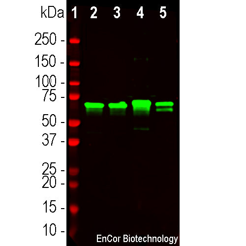
Neurofilaments are the 10nm or intermediate filament proteins found specifically in neurons, and are composed predominantly of three major proteins called NF-L, NF-M and NF-H, though other filament proteins may be included also. The major function of neurofilaments is likely to control the diameter of large axons (1). NF-L is the neurofilament light or low molecular weight polypeptide and runs on SDS-PAGE gels at 68-70kDa with some variability across species. Antibodies to NF-L are useful for identifying neuronal cells and their processes in cell culture and sectioned material. NF-L antibody can also be useful for the visualization of neurofilament rich accumulations seen in many neurological diseases, such as Lou Gehrig’s disease (ALS), giant axon neuropathy, Charcot-Marie-Tooth disease and others (2-4). Much interest has recently been focused on the detection of NF-L released from neurons into blood and CSF as a surrogate marker of primarily axonal loss in a variety of types of CNS injury and degeneration (5).

Chromogenic immunostaining of a formalin fixed paraffin embedded human cerebellum section with rabbit pAb to NF-L, RPCA-NF-L-ct, dilution 1:5,000. detected with DAB (brown) using Vector Labs ImmPRESS method and reagents with citrate buffer retrieval. Hematoxylin (blue) was used as the counterstain. RPCA-NF-L-ct strongly labels the axons and dendrites of Purkinje cells and the projections of neuronal cells within the granular layer. This antibody performs well in testing with both 4% PFA and standard NBF fixed rat, mouse, and human tissues. Mouse select image for larger view.
This antibody is known to cross react well with human, pig and cow NF-L. As shown below the homologous region of the C-terminus of NF-L is not identical to the rodent immunogen used to make RPCA-NF-L-ct but is substantially similar.
Rat NF-L C-terminus GEE-EDTKESEEEEKKEESAGEEQAAKKKD
Identity GEE E+TKE EEEEKK E AGEEQAAKKKD
Human NF-L C-terminus GEEGEETKEAEEEEKKVEGAGEEQAAKKKD
1. Hoffman et al. Neurofilament gene expression:a major determinant of axonal caliber. PNAS 84:3472-6 (1987).
2. Perrot R, et al. Review of the Multiple Aspects of Neurofilament Functions, and their Possible Contribution to Neurodegeneration. Mol. Neurobiol. 38:27-65 (2008).
3. Lépinoux-Chambaud C. Eyer J. Review on intermediate filaments of the nervous system and their pathological alterations. Histochem. Cell Biol. 140:13-22 (2013).
4. Liu Q. et al. Neurofilamentopathy in Neurodegenerative Diseases. Open Neurol. J. 5:58–62 (2011).
5. Bacioglu M, et al. Neurofilament light chain in blood and CSF as marker of disease progression in mouse models and in neurodegenerative diseases. Neuron 91:56-66 (2016).
Add a short description for this tabbed section


%20%20C-terminus,%20Cat%23%20RPCA-NF-L-ct)