

EnCor Biotechnology
Mouse Monoclonal Antibody to β-Synuclein, Cat# MCA-6A10
Description
The MCA-6A10 antibody was made against the C-terminal peptide of human β-synuclein coupled to a carrier protein, and recognizes full length human and rodent β-synuclein specifically both in western blots and in immunocytochemical experiments. The antibody works well for IHC of rodent and human specimens and is completely specific for β-synuclein and does not bind α or γ-synuclein, see these data under the "Additional Data" tab. EnCor also provides a rabbit polyclonal to β-synuclein, RPCA-SCNB, and high quality antibodies to α-synuclein MCA-2A7 and CPCA-SCNA.
- Cell Structure Marker
- Epitope Mapped Antibodies
- Immunohistochemistry Verified
- Mouse Monoclonal Antibodies
- Pathology Related Marker
Add a short description for this tabbed section
| Immunogen: | C-terminal peptide of human β-synuclein EPEGESYEDPPQEEYQEYEPEA coupled to KLH |
| HGNC Name: | SNCB |
| UniProt: | Q16143 |
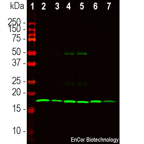
| Molecular Weight: | 17 kDa by SDS-page |
| Host: | Mouse |
| Isotype: | IgG1 |
| Species Cross-Reactivity: | Human, rat, mouse, cow, pig |
| RRID: | AB_2860579 |
| Format: | Protein G affinity purified antibody at 1mg/mL in 50% PBS, 50% glycerol plus 5mM NaN3 |
| Applications: | WB, IF/ICC, IHC |
| Recommended Dilutions: | WB: 1:1,000. IF/ICC: 1:500-1,000. IHC: 1:500-1:1,000. |
| Storage: | Store at 4°C for short term, for longer term store at -20°C. Stable for 12 months from date of receipt. |
β-synuclein is a member of the synuclein protein family, the other two members being α and γ-synuclein, each protein being coded for by a distinct but related gene. α-synuclein was originally isolated as a major synaptic vesicle associated protein from the electric organ of the fish Torpedo (1), and direct homologues of α-synuclein are found in all vertebrates. Later work connected α-synuclein expression with several human brain pathologies, so that it is a major component of the Lewy bodies of Parkinson’s disease (2-5). β-synuclein was isolated as a component of normal and diseased human brain as a protein clearly related to but distinct from α-synuclein (6). The human β-synuclein molecule is 134 amino acids in size compared to 140 amino acids for α-synuclein, and the N-terminal halves of the two molecules are virtually identical while the C-terminal regions is more variable. As a result we made our new β-synuclein antibodies to this region. Like α-synuclein, β-synuclein is heavily concentrated in the brain in presynaptic regions. A third synuclein, γ-synuclein was originally identified as breast cancer specific gene 1, (BCSG1), but is also heavily expressed in brain and also has a similar N-terminal sequence. The three synucleins appear to have overlapping functions so genetic deletion of all three in mice is required to obtain serious neurological deficits (7).


Chromogenic immunostaining of a 4% PFA fixed paraffin embedded rat cerebellum section (top image) and a rat cerebral cortex section (lower image) with mouse mAb to β-synuclein, MCA-6A10, dilution 1:1,000, detected in DAB (brown) following the the Vector Labs ImmPRESS method with citrate buffer retrieval. Hematoxylin (blue) was used as the counterstain. β-synuclein is concentrated in pre-synaptic regions. This antibody performs well in testing with both 4% PFA and standard NBF fixed tissues but does not stain long term NBF fixed tissue effectively. Mouse select image for larger view.

Immunofluorescent analysis of rat cerebellum section stained with mouse mAb to β-synuclein, MCA-6A10, dilution 1:500 in green, and costained with chicken pAb to calbindin, CPCA-Calb, dilution 1:5,000 in red. The blue is Hoechst staining of nuclear DNA. Following transcardial perfusion of rat with 4% paraformaldehyde, brain was post fixed for 24 hours, cut to 45 μM, and free-floating sections were stained with above antibodies. The β-synuclein antibody detects protein concentrated in synaptic regions, and calbindin antibody labels perikarya and dendrites of cerebellar Purkinje cells. Mouse select image at left for larger view.

Western (left) and Ponceau S stained blots (right) of recombinant full length human α, β and γ-synuclein in lanes 2, 3 and 4 respectively. MCA-6A10 reacts strongly with β-synuclein and shows no reaction with the other proteins. Lane 1 shows molecular weight standards of indicated molecular weight. Mouse select image for larger view.
1. Maroteaux L, Campanelli JT, Scheller RH. Synuclein: a neuron-specific protein localized to the nucleus and presynaptic nerve terminal. J. Neurosci. 8:2804-15 (1988).
2. Lavedan C. The Synuclein Family. Genome Research 8:871-80 (1998).
3. Polymeropoulos, MH et al. Mutation in the alpha-synuclein gene identified in families with Parkinson’s disease. Science 276:2045-7 (1997).
4. Kruger, R et al. Ala30-to-Pro mutation in the gene encoding alpha-synuclein in Parkinson’s disease. Nature Genet. 18:106-8 (1998).
5. Chartier-Harlin, M-C. et al. Alpha-synuclein locus duplication as a cause of familial Parkinson’s disease. Lancet 364:1167-9 (2004).
6. Ji H. et al. Identification of a breast cancer-specific gene, BCSG1, by direct differential cDNA sequencing. Cancer Res. 57:759-64 (1997).
7. Greten-Harrison, B. et al. αβγ-Synuclein triple knockout mice reveal age-dependent neuronal dysfunction. PNAS 107:19573-8 (2001).
Add a short description for this tabbed section


