

EnCor Biotechnology
Mouse Monoclonal Antibody to Neurofilament NF-L (Nfl, NEFL), Cat# MCA-1B11
Description
The MCA-1B11 antibody was made against a preparation of NF-L protein purified from pig spinal cord. MCA-1B11 is known to bind NF-L from a variety of species including human, rat and mouse, and the epitope is 100% conserved in all mammalian NF-L sequences, so this antibody will have wide applicability. The epitope is very similar to that of the mouse monoclonal antibody UD1 a.k.a. 47.3, the capture reagent in the NF-Light™ assay, the Quanterix Simoa™ and related NF-L assays. We recently characterized the epitopes for both antibodies used in these assays and developed our own antibodies (6, 7). Interestingly the epitopes are mostly hidden in normal neurofilaments but become accessible on degeneration. Full details of these findings are described in a peer-reviewed publication in Brain Communications. A recent peer reviewed publication shown that this antibody and another EnCor reagent MCA-6H63 can be used to immunoprecipitate proteolytic fragments of NF-L from appropriate human CSF samples (8). We document that MCA-1B11 also works well on paraffin embedded histological sections of rodent CNS tissues, including transgenic mouse models. MCA-1B11 is somewhat "leaky" in that it binds normal neurofilaments when used at higher concentrations but shows strong binding to degenerated material at lower antibody concentrations. For this reason we recommend another mouse monoclonal antibody for the visualization of degenerated NF-L material, MCA-6H63. Another Uman type antibody we market is MCA-1D44. Both MCA-6H63 and MCA-1B11 have been shown to work immunoprecipitate neurodegeneration induced NF-L fragments from human CSF (8). We also market several other NF-L antibodies including a rabbit and chicken polyclonal antibodies RPCA-NF-L-Degen and CPCA-NF-L-Degen and an epitope mapped mouse monoclonals which specifically recognizes non-cleaved froms of NF-L MCA-DA2 and MCA-6H112.
- Cell Structure Marker
- Cell Type Marker
- Cytoskeletal Marker
- Developmental Marker
- Epitope Mapped Antibodies
- Immunohistochemistry Verified
- Mouse Monoclonal Antibodies
- New Antibodies
Add a short description for this tabbed section
| Name: | Neurofilament NF-L, mouse monoclonal, Cat# MCA-1B11 |
| Immunogen: | NF-L purified from pig spinal cord |
| HGNC Name: | NEFL |
| UniProt: | P07196 |
| Molecular Weight: | 68-70kDa |
| Host: | Mouse |
| Isotype: | IgG1 heavy, κ light |
| Species Cross-Reactivity: | Human, rat, mouse, cow, pig, horse |
| RRID: | AB_2737579 |
| Format: | Protein G affinity purified antibody at 1mg/mL in 50% PBS, 50% glycerol plus 5mM NaN3 |
| Applications: | WB, IF/ICC, IHC |
| Recommended Dilutions: | WB: 1:10,000-1:20,000. IF/ICC, and IHC: 1:1,000-1:2,000. |
| Storage: | Store at 4°C for short term, for longer term store at -20°C. Stable for 12 months from date of receipt. |
Neurofilaments are the 10nm or intermediate filament proteins found specifically in neurons, and are composed predominantly of three major proteins called NF-L, NF-M and NF-H, though other filament proteins, but in certain cell types and during development α-internexin, peripherin, nestin and vimentin may be included also. The major function of neurofilaments is likely to control the diameter of large axons (1). NF-L is the neurofilament light or low molecular weight polypeptide and runs on SDS-PAGE gels at 68-70kDa with some variability across species. Antibodies to NF-L are useful for identifying neuronal cells and their processes in cell culture and sectioned material. NF-L antibody can also be useful for the visualization of neurofilament rich accumulations seen in many neurological diseases, such as Lou Gehrig’s disease (ALS), giant axon neuropathy, Charcot-Marie Tooth disease and many others (2-4). Much interest has recently been focused on the detection of NF-L released from neurons into blood and CSF as a surrogate marker of primarily axonal loss in a variety of types of CNS injury and degeneration (5, 6).

Chromogenic Immunostaining of a formalin fixed paraffin embedded brain stem section from a transgenic mouse model of ALS stained with mouse mAb to NF-L, MCA-1B11, dilution 1:1,000, detected with DAB (brown) following the Vector Labs mouse on mouse (MOM) method, without the antigen retrieval step. Hematoxylin (blue) was used as the counterstain. The MCA-1B11 antibody labels what are clearly degenerated axons, showing typical swollen, sinusoidal and discontinuous profiles. Note that under these conditions healthy axons are not stained. Mouse select image for larger view.

This antibody was made against NF-L purified from pig spinal cord and binds to amino acids 316-370 of human NF-L as described in our recent peer reviewed publication in Brain Communications. The MCA-1B11 antibody has an epitope similar to the Uman NF-Light™ assay UD1, also known as 47.3 (7). A shows diagrammatically the location of various NF-L antibody epitopes and B shows peptide sequence of the epitopes for our new antibodies. Mouse select diagram for larger view.

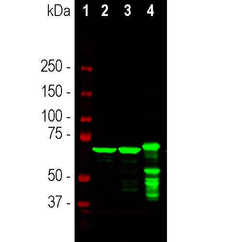
Western blots of Uman NF-LIGHT™ antibodies and a set of EnCor reagents on full length recombinant human PROT-r-NF-L and recombinant human NF-L amino acids 306-364 PROT-r-NF-L-Stan. Lanes labelled 1 in red are protein standards of indicated molecular weights. Lanes labelled 2 were loaded with full length recombinant human NF-L, PROT-r-NF-L, while lanes labelled 3 were loaded with PROT-r-NF-L-Stan. The full length protein runs at about 75kDa, while PROT-r-NF-L-Stan runs at about 12kDa. All five antibodies recognize both constructs. UD1 is also known as 2.1 is the detection reagent in the Uman NF-LIGHT™ assay while UD2, also known as 47.3 is the capture reagent (6). The three other lanes show results obtained with EnCor antibodies MCA-1B11, MCA-4C14 and MCA-6H63 respectively as indicated. All these antibodies binds to an epitope flanking the so-called second "stutter" in the Coil 2 region of the α-helical coiled coil "rod" region of NF-L. The binding properties are very similar to the Uman mouse monoclonal antibody UD1 also known as 2.1, as described originally in Norgren et al. 2002. This antibody is the key capture reagent in the NF-Light™ assay of Uman Diagnostics and the Quanterix Simoa™ NF-L assay. Mouse select image for larger view.
1. Hoffman PN, et al. Neurofilament gene expression:a major determinant of axonal caliber. PNAS 84:3472-6 (1987).
2. Perrot R, etal. Review of the Multiple Aspects of Neurofilament Functions, and their Possible Contribution to Neurodegeneration. Mol. Neurobiol. 38:27-65 (2008).
3. Lépinoux-Chambaud C. and Eyer J. Review on intermediate filaments of the nervous system and their pathological alterations. Histochem. Cell Biol. 140:13-22 (2013).
4. Liu Q, et al. Neurofilamentopathy in Neurodegenerative Diseases. Open Neurol. J. 5:58–62 (2011).
5. Bacioglu M, et al. Neurofilament light chain in blood and CSF as marker of disease progression in mouse models and in neurodegenerative diseases. Neuron 91:56-66 (2016).
6. Norgren N, Karlsson J, E. Rosengren L. and Stigbrand T. Monoclonal antibodies selective for low molecular weight neurofilaments. Hybrid Hybridomics 21:53-59 (2002).
7. Shaw G, et al. Uman type neurofilament light antibodies are effective reagents for the imaging of neurodegeneration. Brain Communications 2023.
8. Becker B, et al. Novel insights into the molecular nature of neurofilament light polypeptide species in cerebrospinal fluid. Brain Communications 2025.
Add a short description for this tabbed section


,%20Cat%23%20MCA-1B11)