

EnCor Biotechnology
Mouse Monoclonal Antibody to Enolase 1 (Non-Neuronal Enolase, NNE, ENO1, α-enolase), Cat# MCA-253
Description
The MCA-253 antibody was made against the N-terminal 12 amino acids of enolase 1 (ENO1), the sequence formed into an 8 armed MAP construct using the procedure of Tam et al. (4). This produces a dendrimer presenting 8 peptides to the immune system obviating the need for coupling to KLH or other carrier protein. This was done by using both the N-terminal and the ε-amino group of lysine for coupling. The specific sequence was therefore (MSILKLVAREIF)8-K4-K2-K.
The antibody works well for western blotting and for IF, ICC and IHC (for IHC see data under "Additional Data" tab). ENO1 is also known as α-enolase or non-neuronal enolase. The production and characterization of this antibody has been described in peer reviewed form, showing specificity of ENO1 and no binding to the related enolases ENO2 and ENO3 (5). We also supply polyclonal antibodies to ENO2, also known as neuron specific enolase (NSE) and γ-enolase, made in rabbit and chicken, RPCA-NSE and CPCA-NSE.
- Cell Type Marker
- Cytoplasmic Enzyme
- Epitope Mapped Antibodies
- Immunohistochemistry Verified
- Mouse Monoclonal Antibodies
Add a short description for this tabbed section
| Immunogen: | N-terminal 12 amino acids of bovine enolase 1 |
| HGNC Name: | ENO1 |
| UniProt: | Q9XSJ4 |
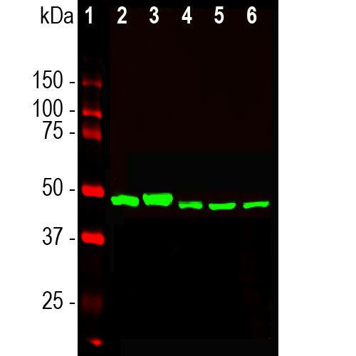
| Molecular Weight: | 47kDa |
| Host: | Mouse |
| Isotype: | IgG1 |
| Species Cross-Reactivity: | Human, rat, mouse, cow, pig, horse |
| RRID: | AB_2572307 |
| Format: | Protein G affinity purified antibody at 1mg/mL in 50% PBS, 50% glycerol plus 5mM NaN3 |
| Applications: | WB, IF/ICC, |
| Recommended Dilutions: | WB: 1:10,000. IF/ICC, and IHC: 1:500-1:1,000. |
| Storage: | Store at 4°C for short term, for longer term store at -20°C. Stable for 12 months from date of receipt. |
Enolase 1 is an enzyme which catalyzes the conversion of 2-phosphoglycerate to phosphoenolpyruvate in the glycolytic pathway, and also the reverse reaction in gluconeogenesis. It is one of three mammalian enolases, which closely are related in protein sequence (see here), and have different cell type specific expression patterns, so that antibodies to them are useful cell type specific markers. Enolase 1 is also known as α enolase and as non-neuronal enolase or NNE. Neuron specific enolase (NSE) corresponds to enolase 2 or γ enolase and is heavily expressed in neuronal cells. The third enolase, enolase 3 or β enolase, is expressed in muscle cells. Enolase 1 is expressed in most kinds of tissue, but is absent from neurons. Abnormal expression of enolase 1 is associated with tumor progression in some breast and head and neck cancer (1,2). We also market antibodies directed against neuronal specific enolase, RPCA-NSE. A switch from enolase 1 to NSE expression occurs in the development of neurons (3).

Chromogenic immunostaining of a 4% PFA fixed paraffin embedded rat cerebellum section with mouse mAb to enolase 1, MCA-253, dilution 1:1,000, detected with DAB (brown) using the the Vector Labs ImmPRESS method and reagents with citrate buffer retrieval. Hematoxylin (blue) was used as the counterstain. The MCA-253 antibody labels the nuclei of non neuronal cells in the molecular and granular cell layers with a concentration at the nuclear membrane. Purkinje and granule neurons are notably negative. This antibody performs well in testing with both 4% PFA and standard NBF fixed human and rat tissues. Mouse select image for larger view.
A sequence alignment of the 3 human enolase molecules can be downloaded from here.
1. Wu W, et al. Identification and validation of metastasis-associated proteins in head and neck cancer cell lines by two-dimensional electrophoresis and mass spectrometry. Clin Exp Metastasis.9(4):319-26 (2002). Clin Exp Metastasis. 19:319-26 (2002).
2. Tu SH, et al. Increased expression of enolase alpha in human breast cancer confers tamifer resistant in human breast cancer cells. Breast Cancer Res. Treat. 121:539-53 (2010).
3. Marangos PJ, Schmechel DE, Parma AM, Goodwin FK. Developmental profile of neuron-specific (NSE) and non-neuronal (NNE) enolase. Brain Res. 190:185-93 (1980).
4. Smith WC, et al. Interaction of arrestin with enolase 1 in photoreceptor. Invest Ophthalmol Vis Sci. 52:1832-40 (2011).
5. Tam JP, Zavala F.J. Multiple antigen peptide. A novel approach to increase detection sensitivity of synthetic peptides in solid-phase immunoassays. J. Immunol Methods. 124:53-61 (1989).
Add a short description for this tabbed section


,%20Cat%23%20MCA-253)