

EnCor Biotechnology
Mouse Monoclonal Antibody to mCherry, Cat# MCA-5A6
Description
The MCA-5A6 antibody was made against full length recombinant mCherry expressed in and purified from E. coli, EnCor product Prot-r-mCherry. MCA-5A6 antibody recognizes mCherry on western blots, in appropriate cells and sections, and does not react with GFP. The antibody has been epitope mapped to within the peptide IKQRLKLKDGGHYDAEVKTT, conserved in mApple and some other fluorescent proteins but not in GFP and others. We have tested this antibody on Tomato, and found that it does not bind. However our alternate mouse monoclonal antibody to mCherry, MCA-1C51, binds equally well to mCherry and Tomato. A sequence alignment of mCherry and GFP with epitope information is here. MCA-5A6 antibody can be used to verify the size of fusion constructs by western blotting, and to amplify the endogenous fluorescence of mCherry in transfected cells. We also supply chicken, CPCA-mCherry and goat polyclonal antibodies to this protein, GPCA-mCherry.
- Epitope Mapped Antibodies
- Fluorescent Protein Antibodies
- Immunohistochemistry Verified
- Mouse Monoclonal Antibodies
Add a short description for this tabbed section
| Immunogen: | Full length recombinant mCherry protein expressed in and purified from E. coli |
| UniProt: | D1MPT3 |
| Molecular Weight: | ~28kDa |
| Host: | Mouse |
| Isotype: | IgG1 heavy, κ light |
| Species Cross-Reactivity: | N.A. |
| RRID: | AB_2861221 |
| Format: | Protein G affinity purified antibody at 1mg/mL in 50% PBS, 50% glycerol plus 5mM NaN3 |
| Applications: | WB, IF/ICC, IHC |
| Recommended Dilutions: | WB: 1:2,000-1:5,000. IF/ICC: 1:500. IHC: 1:2,000-1:5,000. |
| Storage: | Store at 4°C for short term, for longer term store at -20°C. Stable for 12 months from date of receipt. |
The mCherry protein is derived from a natural product, DsRed, originally isolated as a red fluorescent protein from the coral of the genus Discosoma (1). As with other natural fluorescent proteins of Cnidarians (jelly fish, sea anemones and corals), the natural form of the protein forms stable tetramers in vivo. DsRed was engineered to improve its spectral properties and also prevent multimerization in the lab of Roger Tsien, where much work on fluorescent proteins was performed (2). Several further cycles of mutation, directed modification and evolutionary selection produced mCherry, which is monomeric and has an excitation maximum at 587nm and emission maximum at 610nm (3). The protein is widely used as a fluorescent tracer in transfection, transgenic, photobleaching and FRET type experiments. The prototype for these fluorescent proteins is Green Fluorescent Protein (GFP), which is a ~27kDa protein isolated originally from the jellyfish Aequoria victoria (4). The mCherry protein is similar in size and general structural properties to GFP (5,6), but, obviously, produces a red rather than a green fluorochrome.

Chromogenic Immunostaining of formalin fixed paraffin embedded mCherry transfected monkey brain with mouse mAb to mCherry, MCA-5A6, dilution 1:2,000, detected with DAB (brown) following the Vector labs the Vector Labs ImmPRESS method and reagents with citrate buffer retrieval. Hematoxylin (blue) was used as the counterstain. MCA-5A6 specifically detected the soma and axons of mCherry positive neurons in the cerebellum, as expected for this model. Mouse select image for larger view.
This antibody was made against a recombinant construct expressed in and purified from E. coli. The sequence is identical to that found in a series of widely used expression vectors.
1 MVSKGEEDNM AIIKEFMRFK VHMEGSVNGH EFEIEGEGEG RPYEGTQTAK
51 LKVTKGGPLP FAWDILSPQF MYGSKAYVKH PADIPDYLKL SFPEGFKWER
101 VMNFEDGGVV TVTQDSSLQD GEFIYKVKLR GTNFPSDGPV MQKKTMGWEA
151 SSERMYPEDG ALKGEIKQRL KLKDGGHYDA EVKTTYKAKK PVQLPGAYNV
201 NIKLDITSHN EDYTIVEQYE RAEGRHSTGG MDELYK
Epitope mapping of MCA-5A6 was performed using a nested set of 20 amino acid peptides covering the whole sequence and overlapping by 5 amino acids. The peptide IKQRLKLKDGGHYDAEVKTT, shown in red, strongly inhibited binding of MCA-5A6 to mCherry, while the previous and following peptides had no effect suggesting that the central 10 amino acids, KLKDGGHYDA, amino acids 171-180, are a key component of antibody binding.

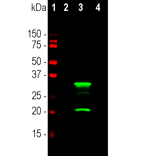
Western blot analysis of HEK293 whole cell lysates using mouse mAb to mCherry, MCA-5A6, dilution 1:2,000, in green [1] protein standard, [2] HEK293, [3] HEK293 cells stably transduced with lentivirus containing mCherry-HA construct, and [4] HEK293 cells transfected with mCherry-HAI construct. Major band at about 28kDa corresponds to mCherry protein. The band at 20kDa corresponds to cleaved form of mCherry protein. Mouse select image for larger view.
1. Matz MV, et al. Fluorescent proteins from nonbioluminescent Anthozoa species. Nat. Biotechnol. 17:969-73 (1999).
2. Baird GS, Zacharias DA, Tsien RY. Biochemistry, mutagenesis, and oligomerization of DsRed, a red fluorescent protein from coral. PNAS 97:11984-9 (2000).
3. Chalfie M, et al. Green fluorescent protein as a marker for gene expression. Science 263:802-5 (1994).
4. Gross LA. et al. The structure of the chromophore within DsRed, a red fluorescent protein from coral. PNAS 97:11990-5 (2000).
5. Heikal AA. et al. Molecular spectroscopy and dynamics of intrinsically fluorescent proteins: coral red (dsRed) and yellow (Citrine). PNAS 97:11996-2001 (2000).
6. Shaner NC. et al. Improved monomeric red, orange and yellow fluorescent proteins derived from Discosoma sp. red fluorescent protein. Nat. Biotech. 22:1567-72 (2004).
Add a short description for this tabbed section


