

EnCor Biotechnology
Mouse Monoclonal Antibody to Neurofilament NF-L (Nfl, NEFL) DegenoTag™ Peptide, Cat# MCA-5H112
Description
MCA-5H112 was raised against a proprietary recombinant immunogen based on the Coil 2 region of human NF-L, the region to which the antibodies described by Norgren et al. bind (7). The antibody works well on western blots of a variety of species but like the Norgren et al. antibodies binds only degenerating or degenerated processes in sectioned material (8). Other Uman type antibodies we market are MCA-1B11 and MCA-6H63. Full details of these findings are described in a peer-reviewed publication in Brain Communications. MCA-5H112 also works on paraffin embedded histological sections of rodent CNS tissues, including transgenic mouse models. It is also an excellent capture reagent in ELISA. EnCor also markets other DegenoTag™ reagents such as MCA-6H63, a mouse monoclonal with a different epitope from MCA-5H112. We also generated degeneration specific chicken and rabbit polyclonals CPCA-NF-L-Degen and RPCA-NF-L-Degen respectively.
- Cell Structure Marker
- Cell Type Marker
- Cytoskeletal Marker
- Epitope Mapped Antibodies
- Immunohistochemistry Verified
- Mouse Monoclonal Antibodies
- New Antibodies
- Pathology Related Marker
Add a short description for this tabbed section
| Immunogen: | Proprietary recombinant construct containing amino acids of human NF-L expressed in and purified from E. coli. |
| HGNC Name: | NEFL |
| UniProt: | P07196 |
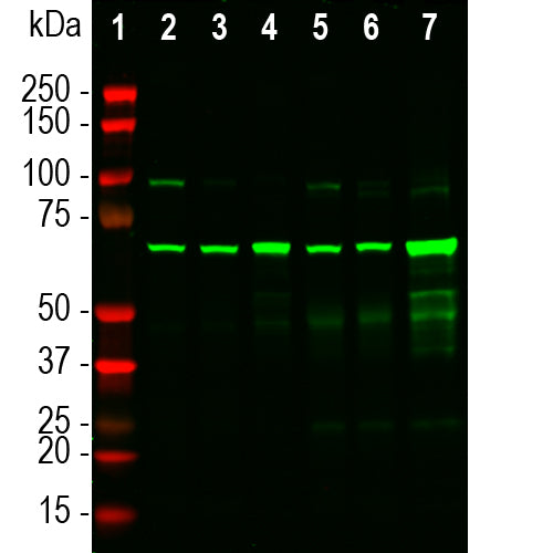
| Molecular Weight: | 68-70kDa by SDS-PAGE |
| Host: | Mouse |
| Isotype: | IgG1 heavy, κ light |
| Species Cross-Reactivity: | Human, rat, mouse, cow, pig |
| RRID: | AB_2923483 |
| Format: | Protein G affinity purified antibody at 1mg/mL in 50% PBS, 50% glycerol plus 5mM NaN3 |
| Applications: | WB, ICC/IF, ELISA |
| Recommended Dilutions: | WB: 1:1,000-1:2,000. ICC/IF, and IHC 1:1,000-1:2,000. |
| Storage: | Store at 4°C for short term, for longer term store at -20°C. Stable for 12 months from date of receipt. |
We have recently developed a series of novel antibody reagents which we call DegenoTag™ products. These are antibodies which recognize epitopes in a small segment of the neurofilament NF-L subunit which are normally not accessible to antibodies but which became available on degeneration. We have evidence that these epitopes are made accessible as a result of degeneration induced proteolysis, and in agreement with this hypothesis we could make previously negative control tissues become strongly DegenoTag™ antibody positive by treatment with proteases. In addition healthy CNS tissues do not stain with DegenoTag™ reagents except for a tiny minority of apparently spontaneously degenerating neuronal cells and processes. In stark contrast DegenoTag™ reagents strongly bind numerous profiles in tissues from animals given experimental spinal cord injuries. We also discovered that our antibodies to the C-terminal of NF-L, such as our rabbit polyclonal RPCA-NF-L-ct and mouse monoclonal MCA-DA2 fail to stain these degenerated profiles. Our reagents can therefore be used to positively identify both healthy and degenerated processes. Process and cells undergoing degeneration show both types of DegenoTag™ reagent.

Chromogenic immunohistochemistry of a 4% PFA fixed paraffin embedded rat spinal cord section taken 3 days after experimental C3 spinal cord injury, resulting in damage to sensory and motor axons. The section was stained with mouse mAb to neurofilament NF-L Degenotag™ antibody MCA-5H112, dilution 1:10,000, detected with DAB (brown) using the Vector Labs ImmPRESS method and reagents with citra buffer retrieval. The MCA-5H112 antibody binds an epitope on NF-L inaccessible to antibodies in healthy axons but exposed in degenerating and degenerated axons. The image shows significant axonal damage in the dorsal columns, containing sensory axons.

Binding of MCA-5H112 to full length recombinant human NF-L in an ELISA in the presence of the indicated 25 amino acid peptides based on the human NF-L sequence. Experiment performed as outlined in our publication in our recent publication in Brain Communications. Peptides 6 to 22 show strong inhibition of binding indicating that the sequence EKQLQELED is a major component of the MCA-5H112 epitope. This sequence is highlighted in red and corresponds to amino acids 330-338 of the human protein. The last 4 lanes labelled Cnt are controls, incubated with an irrelevent peptide.
.
1. Hoffman et al. Neurofilament gene expression:a major determinant of axonal caliber. PNAS 84:3472-6 (1987)
2. Perrot R, et al. Review of the Multiple Aspects of Neurofilament Functions, and their Possible Contribution to Neurodegeneration. Mol. Neurobiol. 38:27-65 (2008).
3. Lépinoux-Chambaud C. Eyer J. Review on intermediate filaments of the nervous system and their pathological alterations. Histochem. Cell Biol. 140:13-22 (2013).
4. Liu Q. et al. Neurofilamentopathy in Neurodegenerative Diseases. Open Neurol. J. 5:58–62 (2011).
5. Bacioglu M, et al. Neurofilament light chain in blood and CSF as marker of disease progression in mouse models and in neurodegenerative diseases. Neuron 91:56-66 (2016).
6. Ayers J. L. et al. Prion-like propagation of mutant SOD1 misfolding and motor neuron disease spread along neuroanatomical pathways. Acta Neuropathol. 131:103-114 (2016).
7. Norgren N, Karlsson JE, Rosengren L, Stigbrand T. Monoclonal antibodies selective for low molecular weight neurofilaments. Hybridoma and Hybridomics 21:53-9 (2002).
8. Shaw G, et al. Uman type neurofilament light antibodies are effective reagents for the imaging of neurodegeneration. Brain Communications doi.org/10.1093/braincomms/fcad067.
Add a short description for this tabbed section


%20DegenoTag%26trade;%20Peptide,%20Cat%23%20MCA-5H112)