
EnCor Biotechnology
Rabbit Polyclonal Antibody to α-Internexin/NF66, Cat# RPCA-a-Int
Description
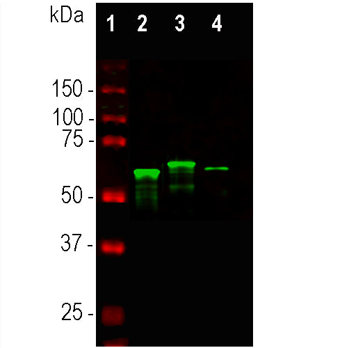
This antibody was made against full length recombinant rat α-internexin fused to the C-terminus of bacterial TrpE expressed in and purified from E. coli. The antibody binds to the α-internexin protein from different mammals, including human, rat, and mouse. It is clean and specific on western blots, ICC and IHC. We also supply mouse monoclonal antibodies, MCA-2E3 and MCA-1D2, and a chicken polyclonal antibody CPCA-a-Int to this protein.
- Cell Structure Marker
- Cell Type Marker
- Cytoskeletal Marker
- Developmental Marker
- Immunohistochemistry Verified
- Pathology Related Marker
- Rabbit Polyclonal Antibodies
Add a short description for this tabbed section
| Immunogen: | Full length recombinant rat α-internexin expressed in and purified from E. coli. |
| HGNC Name: | INA |
| UniProt: | Q16352 |
| Molecular Weight: | 64-66 kDa by SDS-PAGE |
| Host: | Rabbit |
| Species Cross-Reactivity: | Human, rat, mouse, cow, pig, horse |
| RRID: | AB_2572336 |
| Format: | Antibody is supplied as an aliquot of serum plus 5mM NaN3 |
| Applications: | WB, ICC/IF, IHC |
| Recommended Dilutions: | Western blot: 1:10,000. ICC/IF and IHC: 1:500-1:2,000. |
| Storage: | Store at 4°C for short term, for longer term store at -20°C. Stable for 12 months from date of receipt. |
α-internexin is a Class IV intermediate filament protein originally discovered by two different groups of researchers as it copurifies with NF-L, NF-M and NF-H, the then better known major neurofilament "triplet" subunits (1,2). It is expressed only in neurons and in large amounts early in neuronal development, but is down-regulated in many neurons as development proceeds. Some neurons express α-internexin in the absence of NF-L, NF-M and NF-H, though most mature neurons express all four proteins. An α-internexin antibody has been shown, in peer reviewed publications, to reveal the upregulation of α-internexin in facial neurons following experimental axotomy followed by down regulation on axonal regeneration (3). The MCA-2E3 mouse monoclonal antibody to α-internexin, also made by EnCor is the standard reagent used to identify and classify patients with neurofilament inclusion body disease, a specific form of frontotemporal lobar dementia (4-6).

Chromogenic Immunostaining of a formalin fixed paraffin embedded human cerebellum section with rabbit pAb to α internexin, RPCA-a-Int, dilution 1:1,000, detected with DAB (brown) using the the Vector Labs ImmPRESS method and reagents with citrate buffer retrieval. Hematoxylin (blue) was used as the counterstain. The RPCA-a-Int antibody labels axons and dendrites of neuronal cells, outlining the Purkinje cells and parallel fibers in this image. This antibody performs well in testing with both 4% PFA and standard NBF fixed rat, mouse, and human tissues. Mouse select image for larger view.
1. Pachter J and Liem RKH. Alpha-Internexin, a 66-kD intermediate filament-binding protein from mammalian central nervous tissues. J Cell Biol 101:1316-22 (1985).
2. Chiu FC, et al. Characterization of a novel 66 kd subunit of mammalian neurofilaments. Neuron 2:1435-45 (1989).
3. McGraw T. et al. Axonally transported peripheral signals regulate alpha-internexin expression in regenerating motoneurons. J Neurosci. 22:4955-63 (2002).
4. Evans J. et al. Characterization of mitotic neurons derived from adult rat hypothalamus and brain stem. J. Neurophysiol. 87:1076-85 (2002).
5. Cairns NJ. et al. Alpha-internexin is present in the pathological inclusions of neuronal intermediate filament inclusion disease. Am . J. Pathol. 164:2153-61 (2004).
6. Uchikado H1, Shaw G, Wang DS, Dickson DW. Screening for neurofilament inclusion disease using alpha-internexin immunohistochemistry. Neurology 64:1658-9 (2005).
Add a short description for this tabbed section


