

EnCor Biotechnology
Rabbit Polyclonal Antibody to Annexin A6, Cat# RPCA-ANXA6
Description
The RPCA-ANXA6 antibody was made against full length recombinant human annexin A6 expressed in and purified from E. coli. The antibody recognizes annexin A6 in in human, rodents and many other mammals and can be used as a marker of blebs, indicative of cellular stress. We also market a mouse monoclonal antibody to annexin A6 MCA-4G3.
Add a short description for this tabbed section
| Immunogen: | Full length human annexin A6 expressed in and purified from E. coli. |
| HGNC Name: | ANXA6 |
| UniProt: | P08133 |
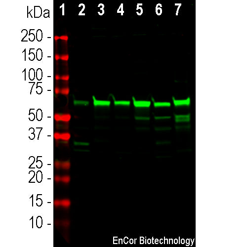
| Molecular Weight: | 75kDa, ~68kDa by SDS-PAGE |
| Host: | Rabbit |
| Species Cross-Reactivity: | Human, Rat, Mouse, Dog, Horse |
| RRID: | AB_3083049 |
| Format: | Immunogen affinity purified antibody at 1mg/mL in 50% PBS, 50% glycerol plus 5mM NaN3 |
| Applications: | WB, IF/ICC, IHC |
| Recommended Dilutions: | WB: 1:5,000. IF/ICC:1:1,000. IHC: 1:2,000. |
| Storage: | Store at 4°C for short term, for longer term store at -20°C. Stable for 12 months from date of receipt. |
The annexins are a large family of related proteins which share the property of binding to phophospholipid containing membranes in a Calcium dependent manner (1). Different members of the family were discovered by different laboratories and as a result the various members have many alternate names, such as lipocortin, calpactin, calelectrin and very many others. In fact Annexin A6 has a particularly surprising number of alternate names, 19 being listed on the Genecards ANXA6 site. The widely used current nomenclature is now based on a letter to indicate membership in a particular one of several annexin sub-families and a number for individual gene products, hence the name annexin A6. The annexin family is defined by a compact disc structure formed from 16 closely packed α-helices which co-ordinate multiple Calcium ions with phospholipid containing membranes. This domain is defined by 4 imperfect repeats of a ~77 amino acid sequence, each repeat forming 4 α-helices (2,3). Annexin A6 was first cloned and sequenced as p68, as a Calcium dependent membrane binding protein extracted from B lymphoblastoid cells (4) and independently as the human homolog of calelectrin, a protein isolated from the electric organ of the fish Torpedo (5). The annexin A6 protein sequence was found to be 75kDa in molecular size, about twice the size of most other annexin family members, and proved to have two of the 16 α-helical regions apparently generated by duplication of the annexin core. The protein has frequently been reported to run on SDS-PAGE more rapidly that expected from the expected 75kDa, at ~68kDa, likely related to the high content of negatively charged amino acids (5). Annexin A6 is normally localized in the cytosol but becomes membrane associated following cellular injury, and so is often seen in ""blebs", transient herniations of the plasma membrane associated with apoptosis and more generally indicative of cell stress. Recent studies suggest that annexin A6 functions, in combination with annexin A4, in the repair of cell membranes following cellular injury (6).

Chromogenic immunostaining of a 4% PFA fixed paraffin embedded rat kidney section with rabbit pAb to annexin A6, RPCA-ANXA6, dilution 1:2,000 detected with DAB (brown) using the Vector Labs ImmPRESS method and reagents with citrate buffer retrieval. Hematoxylin (blue) was used as the counterstain. In this image, RPCA-ANXA6 strongly labels the cytoplasm and membranes of tubule cells. This antibody performs well in testing with both 4% PFA and standard NBF fixed rat, mouse, and human tissues. Mouse select image for larger view.

Chromogenic immunostaining of a 4% PFA fixed paraffin embedded mouse kidney section with rabbit pAb to annexin A6, RPCA-ANXA6, dilution 1:2,000 detected with DAB (brown) using the Vector Labs ImmPRESS method and reagents with citrate buffer retrieval. Hematoxylin (blue) was used as the counterstain. In kidney, RPCA-ANXA6 strongly labels the cytosol of tubule cells, glomeruli and fibroblasts, predominantly in the plasma membrane and endosomal compartments. This antibody performs well in testing with both 4% PFA and standard NBF fixed rat, mouse, and human tissues. Mouse select image for larger view.
1. Gerke V, and Moss SE. Annexins: from structure to function. Physiol Revs 82:331-71 (2002).
2. Barton GJ. et al. Amino acid sequence analysis of the annexin super-gene family of proteins. Eur J Biochem 198:749-60 (1991).
3. Geisow MJ. et al. A consensus amino-acid sequence repeat in Torpedo and mammalian Ca2+-dependent membrane-binding proteins. Nature 320:636-8 (1986).
4, Crompton MR et al. Primary structure of the human, membrane-associated Ca2+-binding protein p68: a novel member of a protein family. EMBO J 7:21-27 (1988).
5. Suedhof TC et al. Human 67-kDa calelectrin contains a duplication of four repeats found in 35-kDa lipocortins. PNAS 85:664-668 (1988).
6. Boye TL et al. Annexin A4 and A6 induce membrane curvature and constriction during cell membrane repair. Nature Comm 8:1623 (2017).
Add a short description for this tabbed section


