


EnCor Biotechnology
Rabbit Polyclonal Antibody to Glial Fibrillary Acidic Protein (GFAP), Cat# RPCA-GFAP
Description
The immunogen used to generate RPCA-GFAP antibody was full length recombinant human GFAP, Prot-r-GFAP, expressed in and purified from E. coli. We document that the antibody works well not only for western blotting, IF and ICC but also on formalin fixed paraffin embedded sections of human and rodent tissues, select the "Additional Data" tab to view this data. The same GFAP immunogen was used to produce chicken, CPCA-GFAP, and goat, GPCA-GFAP polyclonal antibodies. Using different immunogens, EnCor manufactures widely used mouse monoclonal antibodies to GFAP, MCA-5C10, MCA-2A5, and MCA-3E10.
- Cell Structure Marker
- Cell Type Marker
- Cytoskeletal Marker
- Developmental Marker
- Immunohistochemistry Verified
- Our Most Widely Used Reagents
- Pathology Related Marker
- Rabbit Polyclonal Antibodies
Add a short description for this tabbed section
| Immunogen: | Recombinant full length human GFAP isotype 1 expressed in and purified from E. coli. |
| HGNC Name: | GFAP |
| UniProt: | P14136 |
| Molecular Weight: | ~50kDa |
| Host: | Rabbit |
| Species Cross-Reactivity: | Human, rat, mouse, cow, pig, horse |
| RRID: | AB_2572310 |
| Format: | Serum plus 5mM NaN3 |
| Applications: | WB, IF/ICC, IHC |
| Recommended Dilutions: | WB: 1:5,000 IF/ICC and IHC: 1:5,000-1:10,000. |
| Storage: | Store at 4°C for short term, for longer term store at -20°C. Stable for 12 months from date of receipt. |
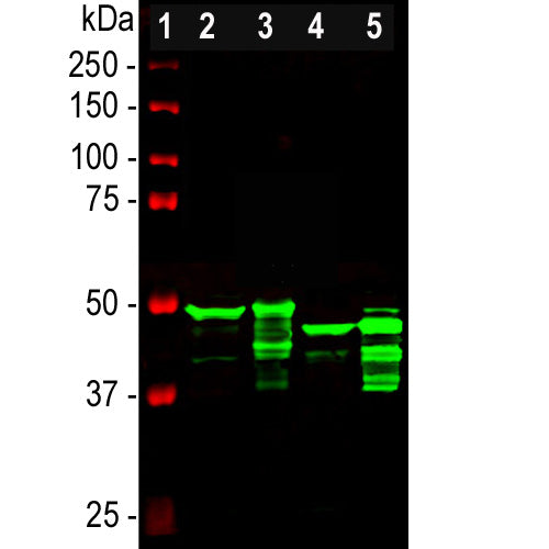
Glial Fibrillary Acidic Protein (GFAP) is a major CNS protein which runs on SDS-PAGE as a ~50kDa protein, usually associated with somewhat lower molecule weight bands which are alternate transcripts from the single gene or in vivo proteolytic fragments. GFAP is strongly and specifically expressed in astrocytes and certain other glia in the central nervous system, in satellite cells in peripheral ganglia, and in non-myelinating Schwann cells in peripheral nerves and is also a component of neural stem cells (1-3). Astrocytes respond to many damage and disease states resulting in “astrogliosis” or the presence of a “glial response”. GFAP antibodies are widely used to see the reactive astrocytes which form part of this response, since reactive astrocytes stain much more strongly with GFAP antibodies than normal astrocytes. GFAP also forms a major component of the so-called glial scar, an astrocyte rich structure apparently forming part of the barrier to nerve fiber regeneration following damage in the central nervous system (4). Neural stem cells frequently strongly express GFAP but many lose this if they develop into neurons or oligodendrocytes. Finally, Alexander disease was recently shown to be caused by point mutations in the protein coding region of the GFAP gene (5). All forms of Alexander disease are characterized by the presence of Rosenthal fibers, which are GFAP containing cytoplasmic inclusions found in astrocytes. Antibodies to GFAP are therefore very useful as markers of glial cells in central and peripheral nerve system, as well as of developing neural stem cells.

Chromogenic immunostaining of a formalin fixed paraffin embedded human cerebellum section with rabbit pAb to GFAP, RPCA-GFAP, dilution 1:5,000, detected with DAB (brown) using the Vector Labs ImmPRESS method and reagents with citrate buffer retrieval. Hematoxylin (blue) was used as the counterstain. The RPCA-GFAP antibody labels the processes of astrocytes within both the granular and molecular layers and Bergmann glia in the molecular layer. This antibody performs well in testing with both 4% PFA and standard NBF fixed rat, mouse, and human tissues. Mouse select image for larger view.

Immunofluorescent analysis of cortical neuron-glial cell culture from E20 rat stained with rabbit polyclonal antibody to GFAP, RPCA-GFAP, dilution 1:5,000 in red, and chicken polyclonal antibody to vimentin, CPCA-VIM, dilution 1:10,000 in green. The blue is DAPI staining of nuclear DNA. The astrocytes containing GFAP only are red, GFAP and vimentin are golden-yellow. Fibroblastic cells contain only vimentin, so they are green. Mouse select image for larger view.

Above is an image generated in the Lifecanvas Technologies laboratory using RPCA-GFAP and an appropriate secondary antibody on a 1mm section of rat brain. This was processed using the Lifecanvas SHIELD fixation protocol which employs a novel epoxy crosslinker (Park et al. Nature Biotechnology 2018). Mouse select image for larger view.
This antibody has become widely used as sold by EnCor and through our numerous OEM partners, and in-formation on this can be viewed using Google scholar by searching for "RPCA-GFAP” or by selecting here. Here is a CiteAb link to peer reviewed publications which use this antibody ob-tained directly from EnCor, here.
1. Bignami A, Eng LF, Dahl D, Uyeda CT. Localization of the glial fibrillary acidic protein in astrocytes by immunofluorescence. Brain Res. 43:429-35 (1972).
2. Yen SH, Fields KL. Antibodies to neurofilament, glial filament, and fibroblast intermediate filament proteins bind to different cell types of the nervous system. J Cell Biol. 88:115-26 (1981).
3. Shaw G, Osborn M, Weber K. An immunofluorescence microscopical study of the neurofilament triplet proteins, vimentin and glial fibrillary acidic protein within the adult rat brain. Eur. J. Cell Biol. 26:68-82 (1981).
4. Fitch MT, Silver J. CNS injury, glial scars, and inflammation: Inhibitory extracellular matrices and regeneration failure. Exp. Neurol. 209:294-301 (2008).
5. Brenner M, et al. Mutations in GFAP, encoding glial fibrillary acidic protein, are associated with Alexander disease. Nat. Genet. 27:117-20 (2001).
The antibody has been sold through many OEM partners, and peer-reviewed publications making use of it can be found by searching Google Scholar for "RPCA-GFAP" or, if you are viewing this online, simply by selecting this link.
Add a short description for this tabbed section


,%20Cat%23%20RPCA-GFAP)