December 2023 News

      We now have new data on two rabbit polyclonal antibodies to microtubule associated protein 2, RPCA-MAP2A/B and a new antibody RPCA-MAP2D. The MAP2 gene produces several different sized proteins by alternate transcription. MAP2C and MAP2D are lower molecular weight isotypes which are expressed early in development and are around 70kDa molecular size when run on SDS-PAGE. MAP2A and MAP2B isotypes are expressed later in development and on SDS-PAGE run at about 220kDa, due to the inclusion of large sequence inserts which produce the MAP2 “projection domains”. Projection domains can be seen in the electron microscope as fine protrusions from the side of microtubules in neuronal perikary and dendrites. Since MAP2C and MAP2D sequences are included in the MAP2A and MAP2B the new RPCA-MAP2D antibody will recognize all forms of MAP2, while RPCA-MAP2A/B will recognize only the mature forms of the protein. Both antibodies work well on western blots, IF, ICC and IHC.
      We went to the Society for Neuroscience Meetimg in Washington DC, 11th to 15th November. Many people visited us at Booth 139 which was easy to remember as it is 30, 31, 32. We gave out the usual pens, posters, flashlights, postcards and antibody samples, and also making available more of our Santiago Ramon-Y-Cajal and Count Camillo Golgi laser cut images.

August 2023 News

      We release yet more novel antibodies. We made a novel rabbit polyclonal and a mouse mpnoclonal to the enzyme catalase, RPCA-Catalase and MCA-6H14, both good markers odf peroxisomes. We also raised rabbit polyclonal antibodies against human Sigma-1 receptor RPCA-Sigma-1r and human γ-synuclein, RPCA-SNCG. We document that all work well on western blots for IF, ICC and IHC.

June 2023 News

We have now finished testing all our rabbit polyclonal antibodies for immunohistochemistry on formalin fixed and paraffin embedded (FFPE) rodent and rodent tissues. So we found that many of them work very well, as shown in the examples below. So now we can recommend (or not) all of our antibodies on FFPE human and rodent specimens, describe the protocols we used and show convincing example images.

Chromogenic immunostaining of a NBF fixed paraffin embedded human brain cortex section with rabbit pAb to IBA1, RPCA-IBA1, dilution 1:2,000, detected with DAB (brown) using the Vector Labs ImmPRESS method and reagents with citra buffer retrieval. Hematoxylin (blue) was used as the counterstain. The RPCA-IBA1 antibody specifically labels the cytoplasm of microglial cells. This antibody performs well in testing with 4% PFA and standard NBF fixed mouse, rat and human tissue.

Chromogenic immunostaining of a formalin fixed paraffin embedded human cerebellum section with rabbit pAb to GFAP, RPCA-GFAP, dilution 1:5,000, detected with DAB (brown) using the Vector Labs ImmPRESS method and reagents with citra buffer retrieval. Hematoxylin (blue) was used as the counterstain. The RPCA-GFAP antibody labels the processes of astrocytes within both the granular and molecular layers and Bergmann glia in the molecular layer. This antibody performs well in testing with both 4% PFA and standard NBF fixed rat, mouse, and human tissues.

Chromogenic immunostaining of a formalin fixed paraffin embedded mouse hippocampus section with rabbit pAb to Fox3/NeuN, RPCA-FOX3, dilution 1:4,000, detected with DAB (brown) using the Vector Labs ImmPRESS method and reagents with citra buffer retrieval. The RPCA-FOX3 antibody selectively labels the nuclei and distal perikaryal of most neuronal cell populations. This antibody performs well in testing with 4% PFA and NBF fixed mouse, human, and rat tissues. Mouse select image for larger view.

Chromogenic immunostaining of a formalin fixed paraffin embedded human cerebellum section with rabbit pAb to NF-L, RPCA-NF-L-ct, dilution 1:5,000, detected with DAB (brown) using the Vector Labs ImmPRESS method and reagents with citra buffer retrieval. Hematoxylin (blue) was used as the counterstain. RPCA-NF-L-ct strongly labels the axons and dendrites of Purkinje cells and the projections of neuronal cells within the granular layer. This antibody performs well in testing with both 4% PFA and standard NBF fixed rat, mouse, and human tissues. Mouse select image for larger view.

Chromogenic immunostaining of a NBF fixed paraffin embedded human midbrain section with rabbit pAb to tyrosine hydroxylase, RPCA-TH, dilution 1:10,000, detected with DAB (brown) using the Vector Labs ImmPRESS method and reagents with citra buffer retrieval. Hematoxylin (blue) was used as the counterstain. In this image, RPCA-TH antibody labels dopaminergic neurons and their axons traversing the striatum. This antibody performs well in testing with 4% PFA and standard NBF fixed mouse, rat and human tissue. Mouse select image for larger view.

March 2023 News

      The data in our recent publication in BioRχiv preprint, entitled “Uman Type NF-L Antibodies Are Effective Reagents for the Imaging of Neurodegeneration” was edited and modified to include further data and was submitted for peer review at the journal “Brain Communications“. It has now been accepted for publication with the same title except that “NF-L” is replaced with “Neurofilament Light” in the title, we added some more data and also another coauthor. An unedited and unformatted version of the paper appeared on-line, see 10.1093/braincomms/fcad067. The same link will access the edited and formatted version of the paper which will be available soon.
      We also add new data for many of our mouse monoclonal antibodies which we have now screen for immunohistochemistry (IHC) on paraffin embedded and formalin fixed human and rodent tissues. For example our widely used mouse monoclonal antibody to c-FOS, MCA-2H2, works well on rat hippocampus highlighting a few active neurons. These cells were expressing large amounts of c-FOS and therefore activating transcription of other genes at the time the tissue was processed and with current techniques can be characterized in terms of mRNA expression and connections. This data is shown under the “Additional Info” tag on the MCA-2H2 web page. Many of our antibodies perform very well and we have posted images under the “Additional Info” tag on the relevant web pages. As expected a few antibodies did not work well and we will add “not recommended for IHC” on the web page, alonfg with details of a n alternate reagent idf we have one. So far we have data on MCA-1B7, MCA-1C51, MCA-2H2, MCA-1C7, MCA-1B11, MCA-1C7, MCA-1C51, MCA-1D2, MCA-1D4, MCA-1C7, MCA-1E3, MCA-1F1, MCA-1H10, MCA-2A7 and MCA-2A8. We are currently screening our entire panel of mouse monoclonal antibodies for utility in IHC, and will shortly post the rest of our results of this.

December 2022 News

      We release two new protein constructs, PROT-r-NF-L-Rct and PROT-r-NF-L-Stan. These constructs are designed to be used as protein standards for current neurofilament NF-L antibody based assays, such as the Uman/Quanterix NF-LIGHT™ and Quanterix Simoa™ assays. The two antibodies used in these assays both bind to the center of the “Coil II” region of α-helical “Rod” region of the NF-L molecule. The PROT-r-NF-L-Rct contains contains the entire Coil II region. The PROT-r-NF-L-Stan constructs contains the epitopes for both antibodies with little flanking sequence and is engineered to include 2 tryptophan residues. The result is a convenient low molecular weight protein standard which due to the high tryptophan content can be quantified spectrophotometrically more accurately than native or recombinant NF-L. Details of the mapping of the relevant NF-L assay antibodies and our generation of a novel panel of similar products are described in a recent BioRχiv publication, 10.1101/2022.08.27.504533v1.
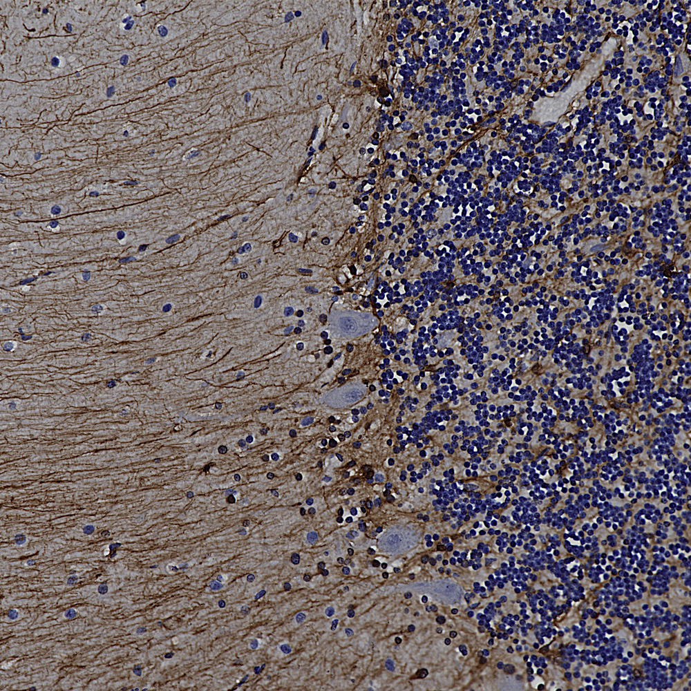
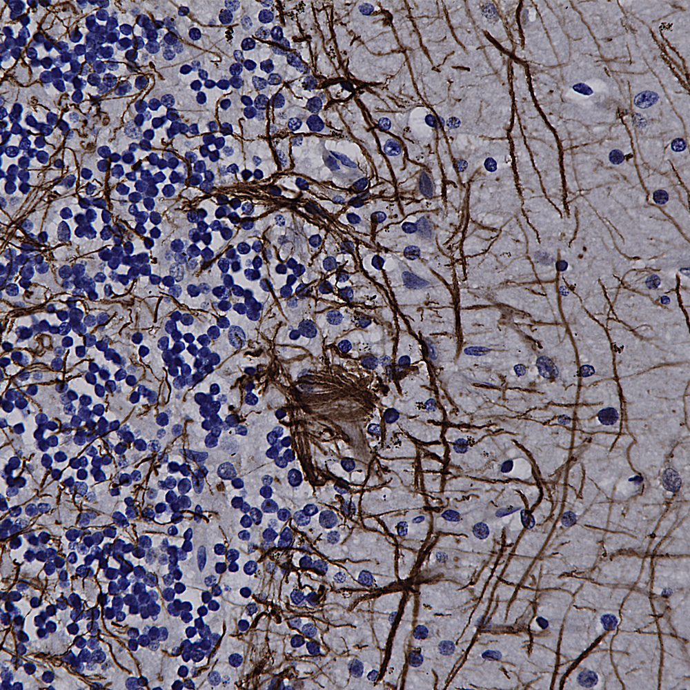
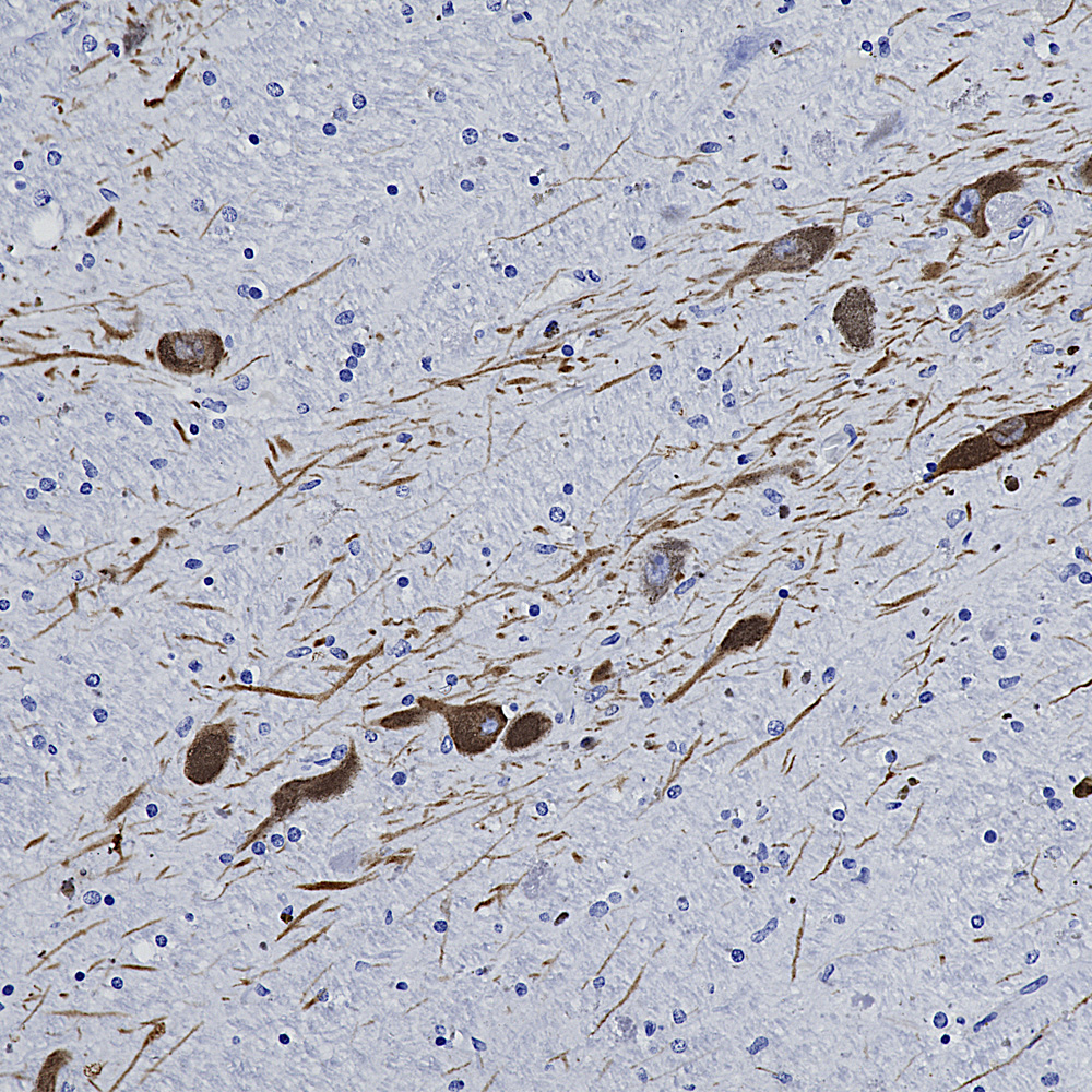
      We hired an excellent immunohistochemist to the great dismay of the University of Florida so we will be validating all our antibodies on formalin fixed paraffin embedded sections, in other words for immunohistochemistry (IHC). The harsh fixation and processing typically used with IHC heavily modifies immunogens and it is therefore expected to cause some otherwise useful antibodies to not work well. However certain other antibodies may recognize epitopes which are not damaged by IHC and so should work just fine. So we added to the “Additional Info” tag of the relevant product web pages images and details of positive IHC results. We now present data on many of our chicken antibodies; Specifically MAP-τ CPCA-Tau, neurofilament NF-H CPCA-NF-H, neurofilament NF-L CPCA-NF-L, tyrosine hydroxylase CPCA-TH, FOX3/NeuN CPCA-FOX3, FOX2 CPCA-FOX2, mCherry CPCA-mCherry, GFP CPCA-GFP, GFAP CPCA-GFAP, IBA1 CPCA-IBA1, MAP2A/B CPCA-MAP2, calbindin CPCA-Calb, myelin basic protein CPCA-MBP, CNPase CPCA-CNP, secretogogin CPCA-SCGN, α-synuclein CPCA-SNCA and visinin-like protein 1 CPCA-VLP1. We have also validated several goat antibodies for formalin fixed paraffin embedded IHC including our antibody to CNPase GPCA-CNP, FOX3/NeuN GPCA-FOX3, GFAP GPCA-GFAP, GFP GPCA-gfP,
MAP2 GPCA-MAP2, MBP GPCA-MBP, mCherry GPCA-mCherry, NF-H GPCA-NF-H and tyrosine hydroxylase GPCA-TH. We have also validated some of our mouse monoclonal antibodies on paraffin embedded IHC, such as DegenoTag™ NF-L antibody MCA-1B11, antibody to mCherry MCA-1C51,
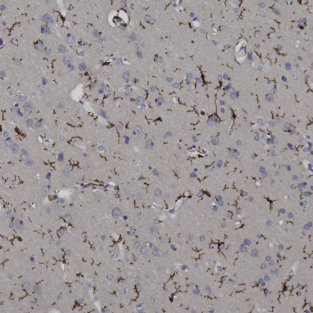
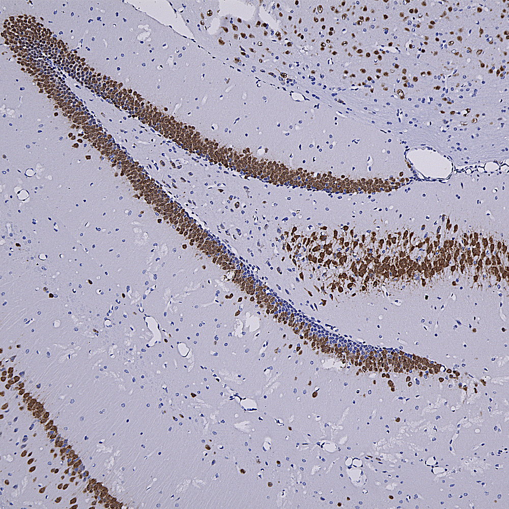
      Over the next few weeks we will validate all of our other chicken, mouse, rabbit and goat antibodies for IHC on human and rodent tissues, significantly enhancing their utility. We have already validated certain of our most widely used monoclonal antibodies such as MCA-1B7 which binds to FOX3/NeuN, an excellent marker of neurons, our rabbit polyclonal antibody to IBA1, RPCA-IBA1 an excellent marker of microglia and our monoclonal antibody to ubiquitin MCA-UBI-1, an excellent marker of Alzheimer neurofibrillary tangles and other pathological inclusions.
      We usually go to the normally annual Society for Neuroscience Meeting, but missed 2020 and 2021 due to the pandemic. This year the meeting was in San Diego, November 11-16, and it was fun to see various people for the first time in three years. We were at booth 401 and handed out ~1,000 of our well known poster images for free, and they had all gone by the middle of the second day. We took along a few of our slides of brain sections and HeLa cell stained with various of antibodies giving 5 distinctly different fluorescence signals at 405nm, 488nm, 541nm, 645nm and 750nm. So on the brain sections it is possible to visualize on one section some combination of nuclei, neuronal cell bodies, neuronal dendrites, myelin sheaths, axons, astrocytes and microglia. In the HeLa cells you could see some combination of nucleus, mitochondria, microfilaments, intermediate filaments, microtubules, plasma membrane, nucleoli and nuclear lamina. It turned out that the various microscope companies loved these, as they had apparently all been looking for such samples to demonstrate how their various 2 photon, confocal, fluorescence etc. microscopes worked. So we are starting to market these as a new line of products. For no particular reason Dr. Shaw also brought along some Ramon-Y-Cajal images which he had burned into wood using a laser and tastefully framed. These turned out to be very popular also, and we sold all of them. So there is yet another potential line of products, watch this space!

August 2022 News

      The Uman Diagnostics NF-LIGHT™ ELISA is a widely used ELISA type assay based on two mouse monoclonal antibodies to the neurofilament NF-L protein. One Uman antibody called variously UD1 or 2.1 is the detection reagent and the other, UD2, also known as 47.3, is the capture reagent (see Norgren et al. 2002). The same pair of antibodies are used in the more sensitive “Single Molecule Array” (Simoa™) assay marketed by Quanterix. These NF-L based assays have become very informative for the quantitation of axonal loss associated with a variety of CNS damage and disease states, see for example Barro et al. 2020 and Gaetani et al. 2019. We now have fully characterized the epitopes for both antibodies and have deposited the basic data on a preprint server see BioRχiv 2022.08.27.504533v1. The data in this report is currently undergoing peer review. These findings greatly increase understanding of what the Uman NF-LIGHT™ ELISA and the Quanterix Simoa™ assay are actually measuring and sets the stage for further NF-L assays with possibly improved properties.
      To our great surprise, neither Uman antibody stained sections of healthy CNS tissue with a typical NF-L pattern. However axons which were damaged as a result of experimental spinal cord injury in rats were strongly reactive with both Uman reagents. We hypothesized that the Uman epitopes were masked in assembled neurofilaments and made available to antibody binding by degeneration induced proteolysis. In agreement with this hypothesis we could make previously Uman negative control tissues strongly Uman positive by treatment with proteases. In addition fresh CNS tissues did not stain with Uman reagents while tissues left to sit at room temperature for 4 hours were strongly reactive. We also discovered that our antibodies to the C-terminal of NF-L, such as our rabbit polyclonal RPCA-NF-L-ct and mouse monoclonal MCA-DA2 stained neurofilaments in healthy processes but generally did not stain the degraded Uman positive NF-L positive material. On closer examination we determined that during injury induced degeneration processes positive for the C-terminal of NF-L became swollen and beaded. They apparently start to express the Uman epitopes and lose the NF-L C-terminal epitopes at about the same time. The final product is mostly diffuse and globular Uman positive degenerated axonal material. The Uman reagents and our NF-L C-terminal antibodies can therefore be used to positively identify both healthy, degenerating and degenerated processes.
      Based on these findings we made a novel panel of antibodies, both monoclonal and polyclonal, to the peptide containing both Uman epitopes. We have named these “DegenoTag™” reagents, as they specifically identify degenerated processes. These have the same properties of the Uman reagents as described above. We therefore have a panel of well characterized antibodies which can be used in the development of ELISA reagents and in immunocytochemical studies of normal, damaged and diseased CNS tissues. The first of these are MCA-6H63, MCA-1B11 and MCA-1D44. MCA-6H63 has an epitope a few amino acids N-terminal to that of UD2 while the epitope for MCA-1D44 is very similar to that of UD2. MCA-1B11 has an epitope similar to that of UD1. MCA-1D44 and MCA-6H63 share the interesting Uman property of only binding to degenerating and degenerated cells and processes, while MCA-1B11 also stains this degenerated material strongly but shows some reactivity with undamaged neurons and processes. We have also developed a rabbit polyclonal antibody RPCA-NF-L-Degen to the same region of NF-L which shares the property of only recognizing forms of NF-L expressed in degenerating processes.
      We publish another research report in collaboration with scientists at the University of Florida. This is “DAT and TH expression marks Human Parkinson’s Disease in Peripheral Immune Cells” by Gopinath et al. from the lab of Habibeh Khoshbouei in the journal “npj Parkinson’s Disease”. This article make use of our tyrosine hydroxylase (TH) antibodies to study blood cells in Parkinson’s disease patients. We also release an excellent and novel goat polyclonal antibody to tyrosine hydroxylase GPCA-TH.

July 2020 News

      Like many other laboratories with expertise with antibody production we have focused some urgent attention on the SARS-Cov2 virus and the ACE2 protein which it uses to enter and infect human cells. The region of the virus spike or S-protein and the corresponding virus binding region on the ACE2 protein were recently characterized so we made recombinant forms of both regions in E. coli (Prot-r-SARS-CoV2-bd and Prot-r-ACE2-bd). We now have a panel of excellent mouse monoclonal antibodies to the ACE2 binding site of the SARS-CoV2 S-protein, and are marketing the first two of these, MCA-5G8 and MCA-2G1. We also made an affinity purified rabbit polyclonal antibody raised against the same immunogen, RPCA-SARS-CoV2-bd. These antibodies can be used to identify the SARS-Cov2 virus on western blots and by immunofluorescence and may also block the binding of the virus to ACE2. Of general interest to the world, the SARS-CoV2 S-protein ACE2 binding region turns out to be an excellent immunogen, so that it is likely an ideal target for vaccine development.
      We release new antibodies to β-synuclein, a mouse monoclonal and a rabbit polyclonal, MCA-6A10 and RPCA-SNCB. β-synuclein, like the closely related α-synuclein, is a major brain protein concentrated in synaptic regions. We also release new antibodies to Annexin A5 MCA-6A12 and RPCA-ANXA5. Annexin A5 is a Calcium and membrane associated protein which is upregulated when cells become apoptotic. We also release two new mouse monoclonals directed against PEA-15 MCA-4D2 and MCA-4D117. This protein was discovered as a “phosphoprotein enriched in astrocytes of 15kDa” (PEA-15) and independently as a “protein enriched in diabetes” (PED) and so is sometimes referred to as PED/PEA-15. PEA-15 is one of the large family of proteins containing a death effector domain (DED) and appears to have important functions in the regulation of cell growth, apoptosis and glucose metabolism. Finally we release a goat polyclonal antibody to myelin basic protein, GPCA-MBP, which can be used to identify and study myelin sheathes, oligodendrocytes and certain types of Schwann cell.