
EnCor Biotechnology
Mouse Monoclonal Antibody to NF-L (Nfl, NEFL) DegenoTag™ Peptide Cat# MCA-1D44
Description
MCA-1D44 was raised against a proprietary recombinant immunogen based on the Coil 2 region of human NF-L, the region to which the antibodies described by Norgren et al. bind (7). The antibody works well on western blots of a variety of species but like the Norgren et al. antibodies binds only degenerating or degenerated processes in sectioned material (8). Other Uman type antibodies we market are MCA-1B11 and MCA-6H63. Full details of these findings are described in a peer-reviewed publication in Brain Communications. It also works well on paraffin embedded histological sections of rodent CNS tissues, including transgenic mouse models. It is also an excellent capture reagent in ELISA. EnCor also markets other DegenoTag™ reagents such as MCA-6H63, a mouse monoclonal with a different neoepitope than MCA-1D44 and the chicken and rabbit polyclonals CPCA-NF-L-Degen and RPCA-NF-L-Degen.
Add a short description for this tabbed section
| Name: | Neurofilament NF-L, mouse monoclonal antibody, Cat# MCA-1D44 |
| Immunogen: | Proprietary recombinant construct containing amino acids of human NF-L expressed in and purified from E. coli. |
| HGNC Name: | NEFL |
| UniProt: | P07196 |
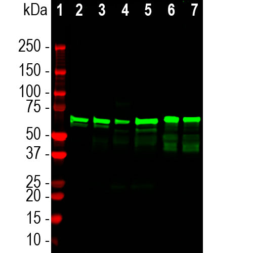
| Molecular Weight: | 68-70kDa by SDS-PAGE |
| Host: | Mouse |
| Isotype: | IgG1 heavy, κ light |
| Species Cross-Reactivity: | Human, rat, mouse, cow, pig |
| RRID: | AB_2923483 |
| Format: | Purified antibody at 1mg/mL in 50% PBS, 50% glycerol plus 5mM NaN3 |
| Applications: | WB, ICC/IF, ELISA |
| Recommended Dilutions: | WB: 1:1,000-1:2,000. ICC/IF, IHC 1:1,000-1:2,000 |
| Storage: | Shipped on ice. Store at 4°C for short term, for longer term at -20°C. Avoid freeze / thaw cycles. |
We have recently developed a series of novel antibody reagents which we call DegenoTag™ products. These are antibodies which recognize epitopes in a small segment of the neurofilament NF-L subunit which are normally not accessible to antibodies but which became available on degeneration. We have evidence that these epitopes are made accessible as a result of degeneration induced proteolysis, and in agreement with this hypothesis we could make previously negative control tissues become strongly DegenoTag™ antibody positive by treatment with proteases. In addition healthy CNS tissues do not stain with DegenoTag™ reagents except for a tiny minority of apparently spontaneously degenerating neuronal cells and processes. In stark contrast DegenoTag™ reagents strongly bind numerous profiles in tissues from animals given experimental spinal cord injuries. We also discovered that our antibodies to the C-terminal of NF-L, such as our rabbit polyclonal RPCA-NF-L-ct and mouse monoclonal MCA-DA2 fail to stain these degenerated profiles. Our reagents can therefore be used to positively identify both healthy and degenerated processes. Process and cells undergoing degeneration show both types of DegenoTag™ reagent.

MCA-1D44 was made against a proprietary immunogen containing amino acids 316-370 as described in our recent peer reviewed publication in Brain Communications. The antibody has an epitope similar to that of the Uman NF-Light™ assay monoclonal UD2, also known as hybridoma 2.1 (7). Above is a diagram showing human NF-L with the location of the epitopes for EnCor and other antibodies. Mouse select diagram for larger view.

Immunohistochemistry on formalin fixed paraffin embedded sections of a mouse model of ALS from the lab of Dr. David Borchelt (6). These heterozygous G85R-SOD1-YFP mice do not develop ALS unless they are primed by injection of mutant SOD1 aggregates, so this is a model of prion induction of pathogenesis. This mouse was injected with mutant SOD1 in the hind limb and degenerated processes were seen in the sciatic nerve, in fibers in the spinal cord and as shown here in the brain stem. The inset shows an example of a helical typical of degenerating processes. Note swollen and sinusoidal profiles. The inset shows an example of a beaded profile typical of degenerating processes. Staining was with MCA-1D44 diluted 1:2,000 detected with DAB (brown) following the Vector Labs mouse on mouse (MOM) method. Mouse select image for larger view.
.
1. Hoffman et al. Neurofilament gene expression:a major determinant of axonal caliber. PNAS 84:3472-6 (1987)
2. Perrot R, et al. Review of the Multiple Aspects of Neurofilament Functions, and their Possible Contribution to Neurodegeneration. Mol. Neurobiol. 38:27-65 (2008).
3. Lépinoux-Chambaud C. Eyer J. Review on intermediate filaments of the nervous system and their pathological alterations. Histochem. Cell Biol. 140:13-22 (2013).
4. Liu Q. et al. Neurofilamentopathy in Neurodegenerative Diseases. Open Neurol. J. 5:58–62 (2011).
5. Bacioglu M, et al. Neurofilament light chain in blood and CSF as marker of disease progression in mouse models and in neurodegenerative diseases. Neuron 91:56-66 (2016).
6. Ayers J. L. et al. Prion-like propagation of mutant SOD1 misfolding and motor neuron disease spread along neuroanatomical pathways. Acta Neuropathol. 131:103-114 (2016).
7. Norgren N, Karlsson JE, Rosengren L, Stigbrand T. Monoclonal antibodies selective for low molecular weight neurofilaments. Hybridoma and Hybridomics 21:53-9 (2002).
8. Shaw G, et al. Uman type neurofilament light antibodies are effective reagents for the imaging of neurodegeneration. Brain Communications doi.org/10.1093/braincomms/fcad067.
Add a short description for this tabbed section


%20DegenoTag%26trade;%20Peptide%20Cat%23%20MCA-1D44)