


EnCor Biotechnology
Chicken Polyclonal Antibody to mCherry, Cat# CPCA-mCherry
Description
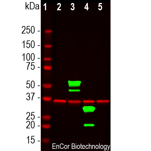
The CPCA-mCherry antibody was made against full length recombinant mCherry protein expressed in and purified from E. coli, prot-r-mCherry, The antibody recognizes mCherry on western blots, in appropriate cells and sections, and does not react with GFP. CPCA-mCherry antibody can be used to verify the size of fusion constructs by western blotting and to amplify the endogenous fluorescence of mCherry in transfected, transduced or transgenic cells. We document that the antibody works well not only for western blotting, IF and ICC but also on formalin fixed paraffin embedded sections, select the "Additional Data" for this data. We also supply mouse monoclonal antibodies to mCherry, MCA-1C51 and MCA-5A6, as well as a goat polyclonal antibody to this protein, GPCA-mCherry .
- Chicken Polyclonal Antibodies
- Fluorescent Protein Antibodies
- Immunohistochemistry Verified
- Our Most Widely Used Reagents
Add a short description for this tabbed section
| Immunogen: | Full length recombinant protein expressed in and purified from E. coli. |
| HGNC Name: | N.A. |
| UniProt: | D1MPT3 |
| Molecular Weight: | ~28kDa |
| Host: | Chicken |
| Species Cross-Reactivity: | Not Applicable |
| RRID: | AB_2572308 |
| Format: | Concentrated IgY preparation in PBS plus 0.02% NaN3 |
| Applications: | WB, IF/ICC, IHC |
| Recommended Dilutions: | WB: 1:2,000-5,000 IC: 1:1000. IF/IHC: 1:5,000-1:10,000. |
| Storage: | Store at 4°C. Stable for 12 months from date of receipt. |
mCherry is derived from proteins originally isolated from Cnidarians (jelly fish, sea anemones and corals), and is used as a fluorescent tracer in transfection and transgenic experiments. The prototype for these fluorescent proteins is green fluorescent protein (GFP), which is a ~27kDa protein isolated originally from the jellyfish Aequoria victoria. GFP was the basis of the 2008 Nobel prize in chemistry, specifically “for the discovery and development of the green fluorescent protein, GFP”. GFP and also mCherry fluoresce on contact with molecular oxygen, requiring no other cofactors, and so can be expressed in fluorescent form in essentially any prokaryotic or eukaryotic cell. The mCherry protein is derived from DsRed, a red fluorescent protein from so-called disc corals of the genus Discosoma. DsRed is similar in size and properties to GFP, but, obviously, produces a red rather than a green fluorochrome. The original DsRed was engineered extensively in the Tsien lab to prevent it from forming tetramers and dimers and to modify and improve the spectral properties. Several further cycles of mutation, directed modification and evolutionary selection produced mCherry, which has an excitation maximum at 587nm and emission maximum at 610nm.The mCherry antibody can used to verify the correct size of mCherry fusions by western blotting in extracts of cells and tissues and to amplify mCherry signals in sectioned material. The same lab engineered other fluorescent DsRed derivatives such as tdTomato, mOrange, mStrawberry and others. This antibody likely binds all these variants and is known to bind tdTomato.
This antibody has become widely used as sold by EnCor and through our numerous OEM partners, and in-formation on this can be viewed using Google scholar by searching for "CPCA-mCherry” or by selecting here. Here is a CiteAb link to peer reviewed publications which use this antibody ob-tained directly from EnCor, here.

Chromogenic immunostaining of formalin fixed paraffin embedded tissue from a mouse transgenically expressing a Cre dependent oxytocin-TdTomato construct in brain tissue. TdTomato is very closely related to mCherry, so CPCA-mCherry would be expected to recognize both proteins. The CPCA-mCherry antibody was used at a dilution 1:10,000, detected with DAB (brown) using the ABC method with citrate buffer retrieval at pH=6.0. Hematoxylin (blue) was used as the counterstain. The CPCA-mCherry specifically detects as expected the soma and axons of Cre expressing neurons. Mouse select image for larger view.
1. Baird GS, Zacharias DA, Tsien RY. Biochemistry, mutagenesis, and oligomerization of DsRed, a red fluorescent protein from coral. PNAS 97:11984-9 (2000).
2. Gross LA, et al. The structure of the chromophore within DsRed, a red fluorescent protein from coral. PNAS 97:11990-5 (2000).
3. Heikal AA, et al. Molecular spectroscopy and dynamics of intrinsically fluorescent proteins: coral red (dsRed) and yellow (Citrine). PNAS 97:11996-2001 (2000).
4. Shaner NC, et al. Improved monomeric red, orange and yellow fluorescent proteins derived from Discosoma sp. red fluorescent protein. Nat. Biotech. 22:1567-72 (2004).
Add a short description for this tabbed section


