

EnCor Biotechnology
Rabbit Polyclonal Antibody to αII-Spectrin, Cat# RPCA-aII-Spec
Description
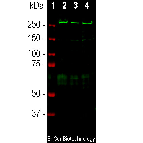
The RPCA-aII-Spec antibody was made against the recombinant human protein construct derived from the C-terminus of αII-spectrin comprising the C-terminal 14 spectrin repeats, specifically amino acids 676-2476. This antibody can be used to study αII-spectrin on western blots and to visualize the neuronal plasma membrane cytoskeleton in cells in culture and sectioned material. We also have made available a mouse monoclonal antibody to the C-terminal region of αII-spectrin which can be used to monitor the presence of calpain and caspase derived spectrin breakdown products MCA-3D7 (7).
- Cell Structure Marker
- Cell Type Marker
- Cytoskeletal Marker
- Immunohistochemistry Verified
- Rabbit Polyclonal Antibodies
Add a short description for this tabbed section
| Immunogen: | Recombinant constructs spanning most of human alpha-II spectrin expressed in and purified from E. coli |
| HGNC Name: | SPTAN1 |
| UniProt: | Q13813 |
| Molecular Weight: | ~240kDa |
| Host: | Rabbit |
| Species Cross-Reactivity: | Human, Rat, Mouse, Cow |
| RRID: | AB_2572382 |
| Format: | Serum plus 5mM NaN3 |
| Applications: | WB, IF/ICC, |
| Recommended Dilutions: | WB: 1:1,000-1:2,000. ICC/IF:500. IHC: Not Recommended . |
| Storage: | Store at 4°C for short term, for longer term store at -20°C. Stable for 12 months from date of receipt. |
Spectrin family molecules are important high molecular weight components of the submembranous cytoskeleton of eukaryotic cells. These proteins were isolated originally from lysed red blood cell membrane preparations which were named "ghosts", which gave rise to the name spectrin (1). Spectrin family molecules are mostly composed of spectrin repeats, compact ~110 amino acid modules made of three closely packed α-helices, though they may also include SH3 domains, PH domains, EF hands and other important binding sites. They function as major components of the membraneous cytoskeleton, mediating interactions between integral membrane proteins, actin and many other cellular components. The RPCA-aII-Spec antibody binds specifically to αII-spectrin, also known as non-erythroid spectrin or fodrin (2-4). In the CNS this protein is expressed only in neurons and so the antibody can be used to reveal the submembranous neuronal cytoskeleton in IF, ICC and IHC. Defects in spectrin genes present as a variety of diseases (5,6). The molecule is subject to proteolysis by calpain producing a 150kDa and 145kDa C-terminal fragments and by caspase producing a slightly different 150kDa C-terminal fragment and a 120kDa C-terminal fragment. Since caspase activation is characteristic of apoptosis and calpain activation of necrosis, it may be possible to use selective monitoring of each type of cell death by monitoring the content of these protein fragments (7).

Chromogenic immunostaining of a formalin fixed paraffin embedded mouse cerebellum section stained with rabbit pAb to αII-spectrin, RPCA- αII-Spec, dilution 1:1,000, detected with DAB (brown) using the Vector Labs ImmPRESS method and reagents with citrate buffer retrieval. Hematoxylin (blue) was used as the counterstain. The RPCA-αII-Spec antibody labels the cytoplasm and membrane of neuronal cells, localizing to vesicles and microtubules. This antibody performs well in testing with both 4% paraformaldehyde and standard neutral buffered saline fixed rat, mouse, and human tissues. Mouse select image for larger view.
1. Marchesi VT, Steers E. Selective solubilization of a protein component of the red cell membrane. Science 159:203-4 (1968).
2. Levine J, Willard M. Fodrin: axonally transported polypeptides associated with the internal periphery of many cells. J. Cell Biol. 90:631-42 (1981).
3. Bennett V, Baines AJ. Spectrin and ankyrin-based pathways: metazoan inventions for integrating cells into tissues. Physiol. Rev. 81:1353-92 (2001).
4. Djinovic-Carugo K, Gautel M, Ylänne J, Young P. The spectrin repeat: a structural platform for cytoskeletal protein assemblies. FEBS Lett. 513:119-23 (2002).
5. Bennett V, Healy J. Organizing the fluid membrane bilayer: diseases linked to spectrin and ankyrin. Trends Mol. Med. 14:28-36 (2008).
6. Eber S, Lux SE. Hereditary spherocytosis–defects in proteins that connect the membrane skeleton to the lipid bilayer. Semin. Hematol. 41:118-41 (2004).
7. Mondello S, et al. αII-spectrin breakdown products (SBDPs): diagnosis and outcome in severe traumatic brain injury patients J. Neurotrauma 27:1203-13 (2010).
Add a short description for this tabbed section


