

EnCor Biotechnology
Chicken Polyclonal Antibody to IBA1 (Allograft Inflammatory Factor 1), Cat# CPCA-IBA1
Description
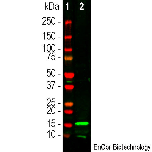
The CPCA-IBA1 antibody was made against the C-terminal peptide of human IBA1 coupled to keyhole limpet hemocyanin and can be used to visualize microglia in the human and rodent brain. We document that the antibody works well not only for western blotting, IF and ICC but also on formalin fixed paraffin embedded IHC sections, select the "Additional Data" for this data. We also market a similar rabbit polyclonal to IBA1 RPCA-IBA1 and also a rabbit antibody to coronin 1a RPCA-Cor1a, another hematopoietic protein specifically expressed in microglia in the nervous system.
- Cell Structure Marker
- Cell Type Marker
- Chicken Polyclonal Antibodies
- Immunohistochemistry Verified
- Pathology Related Marker
Add a short description for this tabbed section
| Immunogen: | Peptide identical to part of the C-terminal of human IBA1 coupled to KLH |
| HGNC Name: | AIF1 |
| UniProt: | P55008 |
| Molecular Weight: | 17kDa |
| Host: | Chicken |
| Species Cross-Reactivity: | Human, rat and mouse |
| RRID: | AB_2923485 |
| Format: | Concentrated IgY preparation plus 0.02% NaN3 |
| Applications: | WB, IF/ICC, IHC |
| Recommended Dilutions: | WB: 1:1,000-5,000. IF 1:1,000-2,000. IHC: 1:5,000-1:10,000. |
| Storage: | Store at 4°C. Stable for 12 months from date of receipt. |
IBA1 is an acronyn for "ionized Calcium binding adapter molecule 1", and the protein is also known as AIF1 for "allograft inflammatory factor 1". AIF1 was originally identified, cloned and sequenced as a protein heavily upregulated in an animal model of graft rejection (1). The AIF1 protein was localized in macrophages and neutrophils surrounding and infiltrating the graft site. Shortly afterwords the same protein was identified a gene product which had some interesting properties, including Calcium binding and the important observation that IBA1 was only expressed in hematopoietic cells (2). IBA1 and AIF1 were subsequently found to be identical, a small globular 17kDa molecule belonging to the "EF" hand superfamily of Calcium binding proteins. Since the only hematopoietic cells and in the neuropil of the central nervous system are microglia, suitable IBA1 antibodies are widely used to identify microglial cells in sections and tissues (3). In tissue samples from which they have not been washed out by perfusion, lymphocytes within blood vessels are also IBA1 positive. Microglia are the immunocompetent cells of the CNS and are extremely important in responses to injury and disease. Microglial are small but very active cells which constantly send processes probing their neighborhood and which alter morphology and are induced to divide following a variety of CNS compromises (4). Many important and highly cited papers have made use of IBA1 antibodies as markers of microglia (e.g. 5,6).

Chromogenic Immunostaining of a formalin fixed paraffin embedded human cerebellum section with chicken pAb to IBA1, CPCA-IBA1, dilution 1:10,000,detected with DAB (brown) following the ABC method with citrate buffer retrieval. Hematoxylin (blue) was used as the counterstain. The IBA1 antibody detects a typical network of microglial processes. Mouse select image for larger view.
1. Utans U, Arceci RJ, Yamashita Y, Russell ME. Cloning and characterization of allograft inflammatory factor-1: a novel macrophage factor identified in rat cardiac allografts with chronic rejection. J. Clin. Invest. 95:2954–62 (1995).
2. Ito D, et al. Microglia-specific localisation of a novel Calcium binding protein, Iba1. Brain Res. Mol. Brain Res. 57:1-9 (1998).
3. Ahmed Z, et al. Actin-binding proteins Coronin-1a and IBA-1 are effective microglial markers for use in Immunohistochemistry. J. Histochem. Cytochem. 55:687-700 (2007).
4. Nimmerjahn A, Kirchhoff F, Helmchen F. Resting Microglial Cells Are Highly Dynamic Surveillants of Brain Parenchyma in Vivo. Science 308:1314-8 (2005).
5. Cardona AE, et al. Control of microglial neurotoxicity by the fractalkine receptor. Nat. Neurosci. 9:917-24 (2006).
6. Mildner A, et al. Microglia in the adult brain arise from Ly-6ChiCCR2+ monocytes only under defined host conditions. Nat. Neurosci. 10:1544-53 (2007).
Add a short description for this tabbed section


,%20Cat%23%20CPCA-IBA1)