

EnCor Biotechnology
Goat Polyclonal Antibody to Myelin Basic Protein (MBP), Cat# GPCA-MBP
Description
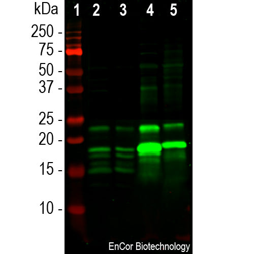
The GPCA-MBP antibody was made against a preparation of MBP purified from bovine brain. It can be used to identify oligodendrocytes and Schwann cells in neural cell culture, to visualize myelin sheaths and myelinating cells in sections and to probe western blots for MBP gene products. The antibody is also rather insensitive to aldehyde fixation and so can be used in immunohistochemistry of formalin fixed paraffin embedded sections. The antibody binds all four of isoforms of MBP on western blots. In contrast our mouse monoclonal MCA-7D2 binds only the 21.5kDa and 18.5kDa rat isotypes, mapping the epitope to the product on one exon, while our MCA-7G7 binds all four isoforms. We also market a chicken polyclonal antibody to MBP CPCA-MBP. A sequence alignment of the four CNS MBP isotypes in human and rat can be downloaded from here.
Add a short description for this tabbed section
| Immunogen: | Purified myelin basic protein isolated from bovine brain |
| HGNC Name: | MBP |
| UniProt: | P02686 |
| Molecular Weight: | 14, 17, 18.5 and 21.5kDa in rodent |
| Host: | Goat |
| Species Cross-Reactivity: | Human, rat, mouse, cow, pig |
| RRID: | AB_2861184 |
| Format: | Immunogen affinity purified antibody at 1mg/mL in 50% PBS, 50% glycerol plus 5mM NaN3 |
| Applications: | WB, IF/ICC, IHC |
| Recommended Dilutions: | WB: 1:5,000. IF/ICC 1:2,000-5,000. IHC 1:5,000-1:10,000. |
| Storage: | Store at 4°C for short term, for longer term at -20°C. Stable for 12 months from date of receipt. |
Myelin Basic Protein (MBP) is one of the major proteins of the myelin sheath surrounding axons in the nervous system. Since it is of relatively low molecular weight and high abundance the protein sequence was determined from purified protein over 30 years ago (1). The protein is made by oligodendrocytes in the central and nervous system, so antibodies to MBP are good markers of this cell type. In the peripheral nervous system MBP is expressed by myelinating Schwann cells so this antibody can be used to identify these cells in culture or sections. In the central nervous system four different forms of the protein made by alternate transcription from a single gene, the protein products having molecular weights of 21.5, 20.5, 18.5, and 17.2kDa in humans. The single gene of rodents also produces 4 different proteins, but of slightly different sizes, 21.5, 18.5, 17 and 14kDa. Some interest has focused on MBP as a potentially significant auto-antigen involved in mouse models of multiple sclerosis (MS, 3) and in human patients (4). Detection of MBP released into blood and CSF has some potential as a surrogate biomarker of demyelination and axonal loss in MS and other relevant damage and disease states (e.g. 5).

Chromogenic immunostaining of a formalin fixed paraffin embedded mouse cerebral cortex section with goat pAb to myelin basic protein (MBP), GPCA-MBP, dilution 1:10,000, detected with DAB (brown) using the ABC method with citrate buffer retrieval at pH=6.0. Hematoxylin (blue) was used as the counterstain. The MBP antibody labels the myelin sheathes around axons network and also oligodendrocyte perikarya. Mouse select image for larger view.

Immunofluorescent analysis of a rat cerebellum section stained with goat pAb to myelin basic protein (MBP), GPCA-MBP, dilution 1:5,000 in red, and costained with rabbit pAb to GFAP, RPCA-GFAP, dilution 1:5,000 in green. The GPCA-MBP antibody stains oligodendrocytes and myelin sheathes around axons, while the GFAP antibody reveals the intermediate filament backbones of astroglial cells. Mouse select image for larger view.
To download a sequence alignment of the 4 human and rat MBP isotypes link to here.
1. Eylar EH, et al. Basic A1 protein of the myelin membrane. The complete amino acid sequence. J. Biol. Chem. 246:5770-84 (1971).
2. Marty MC, et al. The myelin basic protein gene is expressed in differentiated blood cell lineages and in hemopoietic progenitors. PNAS 99:8856-61 (2002).
3. Libbey JE, Fujinami RS. Experimental Autoimmune Encephalomyelitis as a Testing Paradigm for Adjuvants and Vaccines. Vaccine 29:3356–62 (2011).
4. Wucherpfennig KW, Strominger JL. Molecular mimicry in T cell-mediated autoimmunity: Viral peptides activate human T cell clones specific for myelin basic protein. Cell 80:695-705 (1995).
5. Berger RP, et al. Serum neuron-specific enolase, S100B, and myelin basic protein concentrations after inflicted and noninflicted traumatic brain injury in children. J. Neurosurg. 103:61-8 (2005).
Add a short description for this tabbed section


,%20Cat%23%20GPCA-MBP)