
EnCor Biotechnology
Mouse Monoclonal Antibody to Adenylate Cyclase III (ACIII, AC111, AC3), Cat# MCA-1A12
Description
The MCA-1A12 antibody was made against the extreme C-terminal peptide of rat ACIII, PAAFPNGSSVTLPHQVVDNP, amino acids 1125-1144 of the Genbank entry NP_570135.2. A cysteine residue was added to the N-terminus to allow coupling to MBS-activated keyhole limpet hemocyanin. The antibody works on rat and mouse cells which express the same peptide and also on human cells, presumably because the corresponding peptide in the human AC3 sequence is the closely related peptide LATFPNGPSVTLPHQVVDNS. The antibody works well to identify neuronal cilia on both human and rodent cells by IF and ICC but is not recommended for IHC. We have also generated rabbit and chicken polyclonal antibodies to the same ACIII peptide, RPCA-ACIII and CPCA-ACIII.
- Cell Structure Marker
- Cell Type Marker
- Developmental Marker
- Epitope Mapped Antibodies
- Mouse Monoclonal Antibodies
- Not Recommended for IHC
Add a short description for this tabbed section
| Immunogen: | C-terminal peptide of rat ACIII, PAAFPNGSSVTLPHQVVDNP with a Cys added to the N-terminus to allow coupling to KLH. |
| HGNC Name: | ADCY3 |
| UniProt: | P21932 |
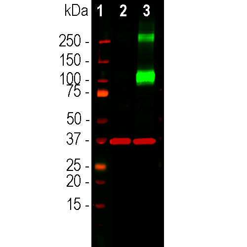
| Molecular Weight: | ~120kDa and above |
| Host: | Mouse |
| Isotype: | IgG1 |
| Species Cross-Reactivity: | Human, Rat, Mouse |
| RRID: | AB_2744501 |
| Format: | Protein G affinity purified antibody at 1mg/mL in 50% PBS, 50% glycerol plus 5mM NaN3 |
| Applications: | WB, ICC |
| Recommended Dilutions: | WB: 1:1,000-1:2,000. ICC: 1:1,000. IF, and IHC: Not Recommended. |
| Storage: | Store at 4°C for short term, for longer term store at -20°C. Stable for 12 months from date of receipt. |
Trimeric G-proteins are a large and variable family of membrane receptors. On binding their specific ligand they activate specific members of the family of trimeric G-proteins which in turn activate other signalling enzymes. Adenylate cyclases are one of these downstream enzyme families which are activated by the GTP bound GαS subunits of trimeric G-proteins. Adenylate cyclases are responsible for the production of the important “second messenger” signaling molecule cyclic-AMP which in turn activates the cAMP dependent protein kinase. This kinase when activated phosphorylates numerous substrate molecules on serine or threonine residues and so alters their activity. There are several different adenylate cyclase genes and protein products with each have distinctly different distribution patterns in cells and tissues. The type III adenylate cyclase enzyme is specifically localized in the membranes surrounding neuronal cilia, and is activated by specific G-protein coupled receptors also located in cilia (1-5). Neuronal cilia express a variety of other receptors types and mediators of other signaling pathways and appear to function as a unique and complex neuronal sensory structure (1-5). For examples, the somatostatin 3 receptor, neuropeptide Y 2 receptor and melanin concentrating hormone receptor 1 are localized in neuronal cilia and the sonic hedgehog and Wnt signalling pathway act on neurons primarily through neuronal cilia (6). This antibody is an excellent marker of neuronal cilia in the brain and in cells in tissue culture and works in the same way as our rabbit polyclonal made against the same peptide (7). The antibody was recently utilized in a very high profile publication in the journal Cell (8).
The antibody has been tested on formalin fixed paraffin embedded human and rodent sections, and is not recommended for this purpose.
1. Fuchs JL, Schwark HD. Neuronal primary cilia: a review. Cell Biol. Int. 28:111-8 (2004).
2. Louvi A and Grove EA. Cilia in the CNS: the quiet organelle claims center stage. Neuron 69:1046-60 (2011).
3. Singla V, Reiter JF. The primary cilium as the cell's antenna: signaling at a sensory organelle. Science 313:629-33 (2006).
4. Green JA, Mykytyn K. Neuronal Primary Cilia: An Underappreciated Signaling and Sensory Organelle in the Brain. Neuropsychopharm. 39:244–5 (2014).
5. May-Simera HL, Kelley MW. Cilia, Wnt signaling, and the cytoskeleton. Cilia 2;1:7 (2012).
6. Guemez-Gamboa A, et al. Primary cilia in the developing and mature brain. Neuron 82:511-21 (2014).
7. Guadiana SM, et al. Arborization of Dendrites by developing neocortical neurons is dependent on primary cilia and Type 3 adenylyl cyclase. J. Neurosci. 33:2626-38 (2013).
8. Sheu S-H et al. A serotonergic axon-cilium synapse drives nuclear signaling to alter chromatin accessibility. Cell 185:3390-3407 (2022).
Add a short description for this tabbed section


,%20Cat%23%20MCA-1A12)