
EnCor Biotechnology
Mouse Monoclonal Antibody to Fibrillarin, Cat# MCA-4A4
Description
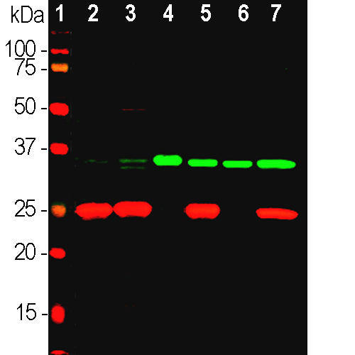
The MCA-4A4 antibody was made against recombinant human fibrillarin expressed in and purified from E. coli and is superior on western blots of mammalian samples to the widely used MCA-38F3 antibody, which was originally raised against yeast Nop1p and later found to recognize fibrillarin, the mammalian homologue of the yeast protein. However MCA-38F3 has been documented to be usable as a marker of nucleoli in a wide variety of species, while MCA-4A4 has only been shown to work on mammalian species. We recently mapped the epitope for MCA-4A4 to TLEPYERDHAVVVGVYRPPP, amino acids 298-317 of the human sequence, at the C-terminal of the globular domain, see here. The KD is 4.22 X 10⁻¹⁰M. We have also produced rabbit and chicken polyclonal antibodies to fibrillarin RPCA-Fib and CPCA-Fib also made against recombinant human fibrillarin.
- Cell Structure Marker
- Epitope Mapped Antibodies
- Immunohistochemistry Verified
- Mouse Monoclonal Antibodies
Add a short description for this tabbed section
| Immunogen: | Recombinant full length human fibrillarin sequence expressed in and purified from E. coli. |
| HGNC Name: | FBL |
| UniProt: | P22087 |
| Molecular Weight: | 34.5kDa |
| Host: | Mouse |
| Isotype: | IgG1 |
| Species Cross-Reactivity: | Human, Rat, Mouse, Horse, Dog, Monkey |
| RRID: | AB_2572264 |
| Format: | Protein G affinity purified antibody at 1mg/mL in 50% PBS, 50% glycerol plus 5mM NaN3 |
| Applications: | WB, IF/ICC, IHC |
| Recommended Dilutions: | WB: 1:2,000. IF/ICC, and IHC: 1:500-1:1,000. |
| Storage: | Store at 4°C for short term, for longer term store at -20°C. Stable for 12 months from date of receipt. |
Fibrillarin is a highly conserved component of a nucleolar small ribonucleoprotein complex in mammals, involved in the processing of ribosomal RNA during ribosomal biogenesis. The protein runs at ~35kDa on SDS-PAGE and is very rich in basic amino acids having a PI of 9.8. Fibrillarin was originally identified in humans since autoantibodies staining nucleoli were seen in some patients with the autoimmune disease scleroderma (1). Subsequently the protein fibrillarin was found to be the human homologue of Nop1p, a Saccharomyces cerevisiae nucleolar protein, the two proteins being 67% identical (2,3). The MCA-38F3 antibody was made against a nuclear preparation from S. cerevisiae and found to bind the yeast protein Nop1p, and was then found to also bind human fibrillarin (2). The fibrillarin molecule consists of an N-terminal glycine and arginine rich region followed by a highly conserved globular domain. Embryonic knockout of the fibrillarin gene in mice is lethal, suggesting fundamental importance of this protein (4). Autoantibodies to fibrillarin are also seen in patients with the autoimmune disease systemic sclerocis (5).

Chromogenic immunostaining of a 4% PFA fixed paraffin embedded rat cerebellum section with mouse mAb to fibrillarin, MCA-4A4, dilution 1:1,000, detected with DAB (brown) using the the Vector Labs ImmPRESS method and reagents with citrate buffer retrieval. Hematoxylin (blue) was used as the counterstain. The MCA-4A4 antibody labels nucleoli most easily observed in cells with a large cytoplasmic to nuclear ratio such as the Purkinje cells in this image. This antibody performs well in testing with both 4% PFA and standard NBF fixed tissues. Mouse select image for larger view.
An alignment of fibrillarin and homologues from a variety of species showing the extremely high cross-species sequence conservation: download from here.
1. Aris JP and Blobel G. Identification and characterization of a yeast nucleolar protein that is similar to a rat liver nucleolar protein. J. Cell Biol. 107:17-31 (1988).
2. Aris JP and Blobel G. cDNA cloning and sequencing of human fibrillarin, a conserved nucleolar protein recognized by autoimmune antisera. Proc. Natl. Acad. Sci. 88:931-5 (1991).
3. Ochs RL, Lischwe MA, Spohn WH, Busch H. Fibrillarin: a new protein of the nucleolus identified by autoimmune sera. Biol. Cell. 54:123-33 (1985).
4. Newton K, Petfalski E, Tollervey D, Caceres JF. Fibrillarin is essential for early development and required for accumulation of an intron-encoded small nucleolar RNA in the mouse. Mol. Cell Biol. 23:8519-27 (2003).
5. Okano Y, Steen VD, Medsger TA. Autoantibody to U3 nucleolar ribonucleoprotein (fibrillarin) in patients with systemic sclerosis. Arth. Rheum. 35:95-100 (1992).
Add a short description for this tabbed section


