


EnCor Biotechnology
Mouse Monoclonal Antibody to Microtubule Associated Protein Tau, MAP-τ (Tau), Cat# MCA-5B10
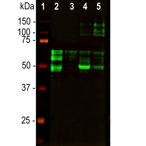
Description
The MCA-5B10 antibody was raised against a recombinant form of one of the lower molecular weight human tau isoform, specifically the human 441 amino acid htau40 form described by Goedert et al. The epitope for this antibody is located in the peptide HVPGGGNKKIETHKLTFREN, amino acids 362-381 of the sequence in NP_005901.2. This corresponds to the C-terminus of the ultimate microtubule binding peptide repeat and some following sequence, see the exact sequence in Tau-Epitopes.pdf. This sequence is expressed in all known human tau isotypes and is totally conserved in all mammals and other higher vertebrates. As a result the antibody will have wide applicability. The antibody works well for western blotting and for IF, ICC and IHC (see data under "Additional Tab" tab). We have another mouse monoclonal antibody raised against the same form of human tau, MCA-2E9, which binds the peptide KDRVQSKIGSLDNITHVPGG, amino acids 347-366, immediately preceding the epitope for MCA-5B10. This is within the ultimate microtubule binding peptide. Both antibodies recognize the non phosphorylated forms of tau. We have not to date examined the effect of tau phosphorylation on binding of either antibody. MCA-5B10 works well on western blots IF, ICC and IHC of human brain tissue.
- Cell Structure Marker
- Cell Type Marker
- Cytoskeletal Marker
- Epitope Mapped Antibodies
- Immunohistochemistry Verified
- Mouse Monoclonal Antibodies
- Pathology Related Marker
Add a short description for this tabbed section
| Immunogen: | Recombinant full length 441 amino acid human tau |
| HGNC Name: | MAPT |
| UniProt: | P10636 |
| Molecular Weight: | 48-67 kDa, with higher molecular weight "big tau" isoforms mostly in peripheral tissues. |
| Host: | Mouse |
| Isotype: | IgG1 |
| Species Cross-Reactivity: | Human, Rat, Mouse |
| RRID: | AB_2572350 |
| Format: | Protein G affinity purified antibody at 1mg/mL in 50% PBS, 50% glycerol plus 5mM NaN3 |
| Applications: | WB, ICC/IF, IHC |
| Recommended Dilutions: | WB: 1:2,000. IF/ICC, and IHC: 1:1,000- 1:2,000. |
| Storage: | Store at 4°C for short term, for longer term store at -20°C. Stable for 12 months from date of receipt. |
MAP-τ is a low molecular weight member of the microtubule associated protein or MAP family. Several serious human diseases are associated with accumulations of MAP-τ, usually referred to as tau protein, most notably the neurofibrillary tangles of Alzheimer's disease. Accumulations of tau in neurons are also characteristic of chronic traumatic encephalopathy, Pick's disease and several other neurodegenerative diseases. Together these disorders are known as "tauopathies". The single mammalian tau gene produces at least 9 different proteins by alternate transcription. In the central nervous system 6 isoforms ranging from 48-67kDa by SDS-PAGE predominate, though larger isotypes are seen mostly in the peripheral nervous system. The tau molecules are very heavily charged and run on SDS-PAGE much more slowly than predicted from their real molecular size. For example the smallest human tau isotype runs at 48kDA on SDS-PAGE but the real molecular weight is 32kDa. Tau proteins are substrates for ser/thr phosphorylation and other post-translational modifications.

Chromogenic Immunostaining of a NBF fixed paraffin embedded human hippocampal section from an Alzheimer’s disease case. Mouse mAb to MAPtau, MCA-5B10, dilution 1:1,000, detected with DAB (brown) using the Vector Labs ImmPRESS method and reagents with citrate buffer retrieval. Hematoxylin (blue) was used as the counterstain. The MCA-5B10 antibody strongly labels nuclei and cell bodies in diseased neurons as identified by their morphology. This antibody performs well in testing with 4% PFA or standard NBF fixed human and rat tissues. Mouse select image for larger view.
This antibody and also our alternate MAP-τ antibody MCA-2E9 was made against recombinant human 3-repeat MAP-τ, the smallest isoform. The immunogen sequence was based on that in NP_005901.2 which was inserted into the eukaryotic expression vector pET30a(+) which adds an N-terminal His-tag and some other sequence, underlined below. This sequence includes a thrombin cleavage site (blue), an S-tag affinity peptide (red) and an enterokinase cleavage site (green).
MHHHHHHSSG LVPRGSGMKE TAAAKFERQH MDSPDLGTDD DDKAMADIGS EFEPRQEFEV 60
MEDHAGTYGL GDRKDQGGYT MHQDQEGDTD AGLKESPLQT PTEDGSEEPG SETSDAKSTP 120
TAEDVTAPLV DEGAPGKQAA AQPHTEIPEG TTAEEAGIGD TPSLEDEAAG HVTQARMVSK 180
SKDGTGSDDK KAKGADGKTK IATPRGAAPP GQKGQANATR IPAKTPPAPK TPPSSGEPPK 240
SGDRSGYSSP GSPGTPGSRS RTPSLPTPPT REPKKVAVVR TPPKSPSSAK SRLQTAPVPM 300
PDLKNVKSKI GSTENLKHQP GGGKVQIINK KLDLSNVQSK CGSKDNIKHV PGGGSVQIVY 360
KPVDLSKVTS KCGSLGNIHH KPGGGQVEVK SEKLDFKDRV QSKIGSLDNI THVPGGGNKK 420
IETHKLTFRE NAKAKTDHGA EIVYKSPVVS GDTSPRHLSN VSSTGSIDMV DSPQLATLAD 480
EVSASLAKQG L 491
Number of amino acids: 491
Molecular weight: 51281.82
Theoretical pI: 6.84
Amino acid composition:
Ala (A) 39 7.9%
Arg (R) 16 3.3%
Asn (N) 11 2.2%
Asp (D) 36 7.3%
Cys (C) 2 0.4%
Gln (Q) 20 4.1%
Glu (E) 29 5.9%
Gly (G) 54 11.0%
His (H) 19 3.9%
Ile (I) 16 3.3%
Leu (L) 23 4.7%
Lys (K) 47 9.6%
Met (M) 10 2.0%
Phe (F) 4 0.8%
Pro (P) 45 9.2%
Ser (S) 50 10.2%
Thr (T) 37 7.5%
Trp (W) 0 0.0%
Tyr (Y) 5 1.0%
Val (V) 28 5.7%
1. Weingarten, M. D., Lockwood, A. H., Hwo, S.-Y. and Kirschner, M. W. A protein factor essential for microtubule assembly. Proc. Nat. Acad. Sci. USA 72:1858–1862 (1975).
2. Cleveland, D. W., Hwo, S. Y., Kirschner, M. W. Purification of tau, a microtubule-associated protein that induces assembly of microtubules from purified tubulin. J Mol Biol. 116:207-25 (1977).
3. Goedert, M., Spillantini, M. G. A century of Alzheimer’s disease. Science 314:777–81 (2006).
4. Ballatore, C., Lee, V. M., Trojanowski, J. Q. Tau-mediated neurodegeneration in Alzheimer’s disease and related disorders. Nat. Rev. Neurosci. 8:663–72 (2007).
5. Wolfe, M. S. Tau mutations in neurodegenerative diseases. J. Biol. Chem. 284:6021-25 (2009).
6. Goedert M, Spillantini MG, Crowther RA. Cloning of a big tau microtubule-associated protein characteristic of the peripheral nervous system. PNAS 89:1983-7 (1992).
7. Bo
yne LJ, Tessler A, Murray M, Fischer I. Distribution of Big tau in the central nervous system of the adult and developing rat. J. Comp. Neurol. 358:279-93 (1995).
8. Goedert M, et al. Multiple Isoforms of human microtubule-associated protein in tau: Sequences and localization in neurofibrillary tangles of Alzheimer's disease. Neuron 3:519-526 (1989).
Add a short description for this tabbed section


,%20Cat%23%20MCA-5B10)