

EnCor Biotechnology
Mouse Monoclonal Antibody to Microtubule Associated Protein MAP2AB (MAP2), Cat# MCA-5H11
Description
This antibody was raised against a preparation of bovine brain MAP2 and the epitope was mapped to the projection domain using a recombinant construct including amino acids 1057-1507 of the human sequence in Prot-r-MAP2-P3. The antibody binds therefore only to the mature MAP2A and MAP2B isoforms and not the MAP2C and MAP2D forms seen early in development. The antibody works well for western blotting and for IF, ICC and IHC (for IHC see data under "Additional Data" tab). This antibody was featured in a recent Youtube video, see here. EnCor markets another monoclonal antibody which binds all MAP2 isoforms, MCA-2C4, and another mouse monoclonal antibody which binds a different epitope in the projection domain of MAP2A and MAP2B, MCA-4H5. EnCor also markets a very popular chicken polyclonal antibody recognizing MAP2A and MAP2B CPCA-MAP2 and also further MAP2 antibodies made in rabbit and goat.
- Cell Structure Marker
- Cell Type Marker
- Cytoskeletal Marker
- Immunohistochemistry Verified
- Mouse Monoclonal Antibodies
Add a short description for this tabbed section
| Immunogen: | Full length purified bovine protein, epitope mapped to projection domain of human sequence, amino acids 1057-1588 using EnCor product Prot-r-MAP2-P3 |
| HGNC Name: | MAP2 |
| UniProt: | P11137 |
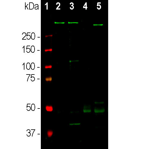
| Molecular Weight: | ~280kDa by SDS-PAGE |
| Host: | Mouse |
| Isotype: | IgG2b heavy, κ light |
| Species Cross-Reactivity: | Human, Rat, Mouse |
| RRID: | AB_2572347 |
| Format: | Protein G affinity purified antibody at 1mg/mL in 50% PBS, 50% glycerol plus 5mM NaN3 |
| Applications: | WB, IF/ICC, IHC |
| Recommended Dilutions: | WB: 1:10,000. IF/ICC: 1:5,000. IHC: 1:5,000-1:10,000. |
| Storage: | Store at 4°C for short term, for longer term store at -20°C. Stable for 12 months from date of receipt. |
Microtubules are 25nm diameter protein rods found in most kinds of eukaryotic cells and are associated with a family of proteins called microtubule associated proteins (MAPs). MAPs play a crucial role in the regulation of microtubule dynamics and interactions in vivo. MAP2 was discovered as a high molecular weight MAP with an SDS-PAGE molecular weight of about 280kDa (1-3). A single mammalian MAP2 gene may generate two high molecular weight proteins of ~280kDa named MAP2A and MAP2B and lower molecular weight forms usually named MAP2C and MAP2D which run on SDS-PAGE gels at 60-70kDa. The 60-70kDa forms are found in neurons early in development, but are later replaced by the higher molecular weight forms (2). The MAP2A and MAP2B forms include a long protein sequence which forms fine filamentous protrusions from the sides of brain microtubules, which is therefore referred to as the projection domain. The epitope for this antibody was mapped to the projection domain so the antibody is specific for MAP2A and MAP2B. This region is one of the prototypes for "intrinsically unstructured regions", a widespread type of protein conformation (4). MAP2 isoforms are expressed only in neurons, specifically in the perikarya and dendrites of these cells. Antibodies to MAP2 isotypes are therefore excellent markers of neuronal dendrites and are useful for identifying neurons in cell culture and sections (e.g. 5-9).

Chromogenic Immunostaining of a formalin fixed paraffin embedded human brain cortex section with mouse mAb to MAP2, MCA-5H11, dilution 1:5,000, detected with DAB (brown) using the Vector Labs ImmPRESS method and reagents with citrate buffer retrieval. Hematoxylin (blue) was used as the counterstain. MCA-5H11 strongly labels neurons, neuronal projections, and synapses. This antibody performs well in testing with 4% PFA fixed or standard NBF fixed rat and human tissue but does not stain long term NBF fixed material effectively. Due to its strong staining profile and ability to stain human tissue, MCA-5H11 is our recommended clone for MAP2 immunostaining. Mouse select image for larger view.

Chromogenic immunostaining of a 4% PFA fixed paraffin embedded rat cerebellum section with mouse mAb to MAP2, MCA-5H11, dilution 1:5,000, detected with DAB (brown) using the Vector Labs ImmPRESS method and reagents with citrate buffer retrieval. Hematoxylin (blue) was used as the counterstain. In the cerebellum, MCA-5H11 strongly labels neurons, neuronal projections, and synapses. This antibody performs well in testing with 4% PFA fixed or standard NBF fixed rat and human tissue but does not stain long term NBF fixed material effectively. Due to its strong staining profile and ability to stain human tissue, MCA-5H11 is our recommended clone for MAP2 immunostaining. Mouse select image for larger view.

Immunofluorescent analysis of a rat cerebellum section stained with mouse mAb to MAP2, MCA-5H11, dilution 1:5,000 in green, and costained with rabbit pAb to α-internexin, RPCA-a-Int, dilution 1:2,000 in red. Following transcardial perfusion of rat with 4% paraformaldehyde, brain was post fixed for 24 hours, cut to 45 μM, and free-floating sections were stained with above antibodies. The MCA-5H11 antibody labels MAP2 protein in the perikarya and dendrites of the most neurons, particularly in granular cell and molecular layers of the cerebellum, while the α-internexin antibody selectively stains axons and dendrites of neuronal cells. Mouse select image for larger view.
1. Dehmelt H, Halpain S. The MAP2/Tau family of microtubule-associated proteins.
Genome Biol. 6:204 (2005).
2. Nunez J. Immature and mature variants of MAP2 and tau proteins and neuronal plasticity. Trends Neurosci. 11:477-9 (1998).
3. Vallee R. A taxol-dependent procedure for the isolation of microtubules and microtubule-associated proteins (MAPs). J. Cell Biol. 92:435-42 (1992).
4. Tompa P. Intrinsically unstructured proteins. Trends Biochem. Sci. 27:527-33 (2002).
5. Goetz AK, et al. Temporally restricted substrate interactions direct fate and specification of neural precursors derived from embryonic stem cells. PNAS 103:11063-8 (2006).
6. Walton, NM, et al. Gliotypic neural stem cells transiently adopt tumorigenic properties during normal differentiation. Stem Cells 27:280-9 (2009).
7. Gasser, A. et al. An ankyrinG-binding motif is necessary and sufficient for targeting Nav1.6 sodium channels to axon initial segments and nodes of Ranvier. J. Neurosci. 32:7232-43 (2012).
8. Rush AM, et al. Differential modulation of sodium channel Nav1.6 by two members of the fibroblast growth factor homologous factor 2 subfamily. Eur. J. Neurosci. 23:2551-62 (2006).
9. Eckenstein FP, McGovern T, Kern D, Deignan J. Neuronal vulnerability in transgenic mice expressing an inducible dominant-negative FGF receptor. Exp. Neurol. 198:338-49 (2006).
Add a short description for this tabbed section


,%20Cat%23%20MCA-5H11)