

EnCor Biotechnology
Mouse Monoclonal Antibody to Neurofilament NF-M (Nfm, NEFM), Cat# MCA-3H11
Description
The MCA-3H11 antibody was made against a recombinant fusion protein of E. coli TrpE fused to the C-terminus of rat NF-M, amino acids 677-845 (7). The epitope was mapped using protein cleavage to the extreme C-terminal sequence, amino acids 762-845, a region of very conserved protein sequence which includes some interesting sequence repeats of currently unknown function (8). This epitope corresponds to amino acids 833-916 of the slightly larger human NF-M sequence. The antibody works on a variety of species and is clean on western blots of crude lysates, on cells in culture and staining of sectioned material, including on IHC of rodent and human material (see under "Additional Data" tab). The antibody has been widely used for many years, for example the original observation that the widely used HEK293 cell line has unexpected neuronal properties was made with this antibody (9). Also available from EnCor are rabbit and chicken polyclonal antibodies to the same immunogen RPCA-NF-M, and CPCA-NF-M. All three antibodies works on a variety of species and are clean and specific on western blots, cell and tissue staining.
- Cell Structure Marker
- Cell Type Marker
- Cytoskeletal Marker
- Epitope Mapped Antibodies
- Immunohistochemistry Verified
- Mouse Monoclonal Antibodies
- Pathology Related Marker
Add a short description for this tabbed section
| Immunogen: | Recombinant fusion protein containing the extreme C-terminus of rat NF-M, amino acids 677-845, expressed in and purified from E. coli |
| HGNC Name: | NEFM |
| UniProt: | P07197 |
| Molecular Weight: | 145-160kDa |
| Host: | Mouse |
| Isotype: | IgG1 |
| Species Cross-Reactivity: | Human, rat, mouse, cow, pig, horse, chicken |
| RRID: | AB_2572365 |
| Format: | Protein G affinity purified antibody at 1mg/mL in 50% PBS, 50% glycerol plus 5mM NaN3 |
| Applications: | WB, IF/ICC, IHC |
| Recommended Dilutions: | WB: 1:5,000-1:10,000. IF/ICC: 1:2,000-1:5,000. IHC: 1:5,000-1:10,000. |
| Storage: | Store at 4°C for short term, for longer term store at -20°C. Stable for 12 months from date of receipt. |
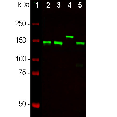
Neurofilaments are the 10nm or intermediate filament proteins found specifically in neurons, and are composed predominantly of three major proteins called NF-L, NF-M and NF-H. NF-M is the neurofilament middle or medium molecular weight polypeptide and runs on SDS-PAGE gels at 145-160kDa, with some species variability, though the real molecular weight is ~105kDa. The major function of neurofilaments is likely to control the diameter of large axons (1). Antibodies to NF-M such as MCA-3H11 are useful for identifying neuronal cells and their processes in tissue sections and in cell culture. NF-M antibodies can also be useful to visualize neurofilament rich accumulations seen in many neurological diseases, such as Amyotrophic Lateral Sclerosis (a.k.a. Lou Gehrig’s disease) and Alzheimer’s disease (2-4). Much recent evidence has suggested that the detection of NF-L and NF-H in blood and CSF might be a useful prognostic or diagnostic biomarkers of neuronal damage and degeneration associated with a variety of CNS pathologies (5,6). The potential utility of NF-M in this fashion has not to date been examined.

Chromogenic immunostaining of a 4% PFA fixed paraffin embedded rat cerebellum section with mouse mAb to NF-M, MCA-3H11, dilution 1:10,000, detected with DAB (brown) using the the Vector Labs ImmPRESS method and reagents with citrate buffer retrieval. Hematoxylin (blue) was used as the counterstain. This antibody performs well in testing with both 4% PFA and standard NBF fixed tissues but does not stain long term NBF fixed tissue effectively. Mouse select image for larger view.
The epitope for MCA-3H11 is located in the C-terminal 84 amino acids of NF-M, a region of highly conserved protein sequence of currently unknown function (7). In the rat sequence this is amino acids 762-845, which corresponds to 833-916 of the human sequence.
See references in peer reviewed publications for the reagent obtained from EnCor here.
1. Hoffman et al. Neurofilament gene expression:a major determinant of axonal caliber. PNAS 84:3472-6 (1987).
2. Perrot R, et al. Review of the Multiple Aspects of Neurofilament Functions, and their Possible Contribution to Neurodegeneration. Mol. Neurobiol. 38:27-65 (2008).
3. Lépinoux-Chambaud C. Eyer J. Review on intermediate filaments of the nervous system and their pathological alterations. Histochem. Cell Biol. 140:13-22 (2013).
4. Liu Q. et al. Neurofilamentopathy in Neurodegenerative Diseases. Open Neurol. J. 5:58–62 (2011).
5. Bacioglu M, et al. Neurofilament light chain in blood and CSF as marker of disease progression in mouse models and in neurodegenerative diseases. Neuron 91:56-66 (2016).
6. Shaw G. The use and potential of pNF-H as a general blood biomarker of axonal loss: an immediate application for CNS injury. in Brain Neurotrauma: Molecular, Neuropsychological, and Rehabilitation Aspects. CRC Press/Taylor & Francis Chapter 21 (2015).
7. Harris J, Ayyub C. and Shaw G. A molecular dissection of the carboxyterminal tails of the major neurofilament subunits NF-M and NF-H. J. Neurosci. Res. 30:47-62 (1991).
8. Shaw G. Identification of previously unrecognized sequence motifs at the extreme carboxyterminus of the neurofilament subunit NF-M. BBRC 14;162:294-9 (1989).
9. Shaw G, et al. Preferential transformation of human neuronal cells by human adenoviruses and the origin of HEK 293 cells. FASEB J. 16:869-71 (2002).
Add a short description for this tabbed section


,%20Cat%23%20MCA-3H11)