

EnCor Biotechnology
Mouse Monoclonal Antibody to Neurofilament NF-L (Nfl, NEFL), Cat# MCA-6H112
Description
MCA-6H112 was made against the peptide GEEEDTKESEEEEKKEESAGEEQVAKKKD with an N-terminal cysteine added by which it was coupled to keyhole limpet hemocyanin. This is the C-terminal peptide of human NF-L, amino acids 514-542, see here. The properties of this antibody make is useful for western blotting, cell and tissue staining and monitoring NF-L proteolysis. The antibody works well for western blotting and for IF, ICC and IHC (for IHC see data under "Additional Data" tab). We also market several other NF-L antibodies including a rabbit and chicken polyclonal antibodies, RPCA-NF-L and a CPCA-NF-L, both made against full length recombinant human NF-L. We also have several epitope mapped mouse monoclonals including the widely used and epitope mapped MCA-DA2, see here.
- Cell Structure Marker
- Cell Type Marker
- Cytoskeletal Marker
- Epitope Mapped Antibodies
- Immunohistochemistry Verified
- Mouse Monoclonal Antibodies
- New Antibodies
- Pathology Related Marker
Add a short description for this tabbed section
| Immunogen: | C-terminal peptide of human NF-L protein, GEEEDTKESEEEEKKEESAGEEQVAKKKD with an N-terminal C for coupling to KLH |
| HGNC Name: | NEFL |
| UniProt: | P07196 |
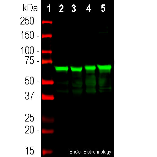
| Molecular Weight: | 68kDa by SDS-PAGE |
| Host: | Mouse |
| Isotype: | IgG1 |
| Species Cross-Reactivity: | Human, Rat, Mouse, Cow, Pig |
| RRID: | AB_2813766 |
| Format: | Protein G affinity purified antibody at 1mg/mL in 50% PBS, 50% glycerol plus 5mM NaN3 |
| Applications: | WB, IF/ICC, IHC |
| Recommended Dilutions: | WB: 1:5,000. IF/ICC: 1:1,000. IHC 1:4,000. |
| Storage: | Store at 4°C for short term, for longer term store at -20°C. Stable for 12 months from date of receipt. |
Neurofilaments are the 10nm or intermediate filament proteins found specifically in neurons, and are composed predominantly of three major proteins called NF-L, NF-M and NF-H, though other filament proteins may be included also. The major function of neurofilaments is likely to control the diameter of large axons (1). NF-L is the neurofilament light or low molecular weight polypeptide and runs on SDS-PAGE gels at 68-70kDa with some variability across species. Antibodies to NF-L like MCA-6H112 are useful for identifying neuronal cells and their processes in cell culture and sectioned material. NF-L antibody can also be useful for the visualization of neurofilament rich accumulations seen in many neurological diseases, such as Lou Gehrig’s disease (ALS), giant axon neuropathy, Charcot-Marie Tooth disease and others (2-4). Much interest has recently been focused on the detection of NF-L released from neurons into blood and CSF as a surrogate marker of primarily axonal loss in a variety of types of CNS injury and degeneration (5).

Chromogenic immunostaining of a formalin fixed paraffin embedded human cerebellum section with mouse mAb to NF-L, MCA-6H112, dilution 1:4,000, detected with DAB (brown) using the the Vector Labs ImmPRESS method and reagents with citrate buffer retrieval. Hematoxylin (blue) was used as the counterstain. MCA-6H112 labels neuronal cells and their processes. This antibody performs well in testing with both 4% PFA and standard NBF fixed tissues and is our recommended clone for use in NF-L immunostaining of human tissue. Mouse select image for larger view.
1. Hoffman et al. Neurofilament gene expression:a major determinant of axonal caliber. PNAS 84:3472-6 (1987).
2. Perrot R, et al. Review of the Multiple Aspects of Neurofilament Functions, and their Possible Contribution to Neurodegeneration. Mol. Neurobiol. 38:27-65 (2008).
3. Lépinoux-Chambaud C. Eyer J. Review on intermediate filaments of the nervous system and their pathological alterations. Histochem. Cell Biol. 140:13-22 (2013).
4. Liu Q. et al. Neurofilamentopathy in Neurodegenerative Diseases. Open Neurol. J. 5:58–62 (2011).
5. Bacioglu M, et al. Neurofilament light chain in blood and CSF as marker of disease progression in mouse models and in neurodegenerative diseases. Neuron 91:56-66 (2016).
Add a short description for this tabbed section


,%20Cat%23%20MCA-6H112)