EnCor Biotechnology
Rabbit Polyclonal Antibody to Heat Shock Protein 60 (HSP60), Cat# RPCA-HSP60
Description
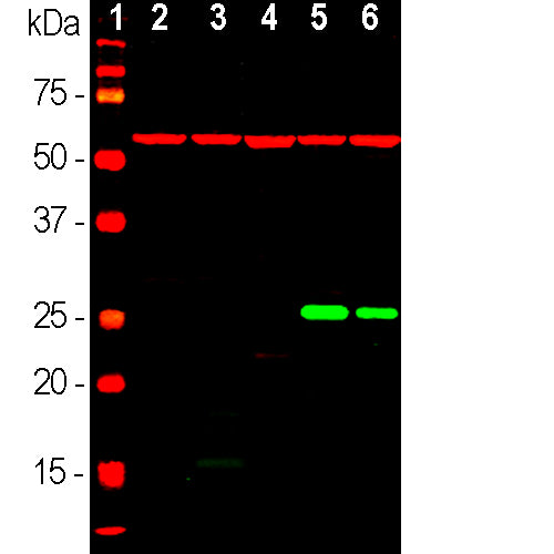
RPCA-HSP60 antibody was made against full length recombinant human HSP60 protein. The same immunogen was also used to generate a chicken polyclonal antibody to HSP60, CPCA-HSP60.These and our mouse monoclonal to HSP60 MCA-1C7 are excellent markers of mitochondria using IF/ICC procedures and recognize HSP60 on western blots producing a single band at 60kDa. This antibody does not perform well for IHC staining of formalin fixed paraffin embedded sections.
Add a short description for this tabbed section
| Immunogen: | Recombinant full length human HSP60 expressed in and purified from E. coli |
| HGNC Name: | HSBD1 |
| UniProt: | P10809 |
| Molecular Weight: | 60kDa |
| Host: | Rabbit |
| Species Cross-Reactivity: | Human, Rat, Mouse, Cow, Pig, Horse, Dog, Monkey |
| RRID: | AB_2572332 |
| Format: | Serum plus 5mM NaN3 |
| Applications: | WB, IF, ICC |
| Recommended Dilutions: | WB: 1:5,000. ICC and IF: 1:1,000. |
| Storage: | Store at 4°C for short term, for longer term store at -20°C. Stable for 12 months from date of receipt. |
The heat shock proteins were discovered, as the name suggests, since they are heavily upregulated when cells are stressed by temperatures above the normal physiological range. They are expressed in unstressed cells also and have a normal function as chaperones, helping other proteins to fold correctly. The need for chaperones is much greater if a cell or tissue is stressed by heat, and so these proteins become heavily up regulated. The different heat shock proteins were originally named based on their SDS-PAGE mobility, so HSP60 has an apparent molecular weight of 60kDa. It is an abundant protein in mitochondria and is typically responsible for the transportation and refolding of proteins from the cytoplasm into the mitochondrial matrix 1,2). In addition to its role as a heat shock protein, HSP60 plays an important role in the transport and maintenance of mitochondrial proteins as well as the transmission and replication of mitochondrial DNA (3,4). HSP60 has been implicated in the initiation and/or progression of some subtypes of cardiovascular disease (CVD), implying its potential as a biomarker with applications for diagnosis, assessing prognosis and response to treatment, as well as for preventing and treating CVD (5). HSP60 appears to be unusually immunogenic, frequently generating autoantibodies in humans and other species (e.g. 6). The HSP60 protein presumably released from damaged or degenerated cells is also a strong inducer of the innate immune system (7).
Our original monoclonal antibody to HSP60
Not Recommended for IHC(p). Please see alternatives CPCA-HSP60 and MCA-1C7.
1. Radford JC, Coates AR, Henderson B. Chaperonins are cell-signalling proteins: the unfolding biology of molecular chaperones. Expert Rev. Mol. Med. 2:1–17 (2000).
2. Bukau B, Horwich AL.The Hsp70 and Hsp60 Chaperone Machines Cell 92:351-66 (2000).
3. Koll H, et al. Antifolding activity of hsp60 couples protein import into the mitochondrial matrix with export to the intermembrane space. Cell 68:1163–75 (1992).
4. Kaufman BA. Kolesar JE, Perlman PS, Butow RA. A function for the mitochondrial chaperonin Hsp60 in the structure and transmission of mitochondrial DNA nucleoids in Saccharomyces cerevisiae. J. Cell Biol. 163:457-61 (2003).
5. Rizzo M, et al. Heat shock protein-60 and risk for cardiovascular disease. Curr. Pharm. Des. 17:3662-8 (2011).
6. Pockley AG, et al. Identification of human heat shock protein 60 (Hsp60) and anti-Hsp60 antibodies in the peripheral circulation of normal individuals. Cell Stress Chaperones 4:29-35 (1999).
7. Kol A, et al. Cutting edge: heat shock protein (HSP) 60 activates the innate immune response: CD14 is an essential receptor for HSP60 activation of mononuclear cells. J. Immunol. 164:13-7 (2000).
Add a short description for this tabbed section


