| Name: | Mouse monoclonal antibody to GFP |
| Immunogen: | The prot-r-AcGFP recombinant protein purified from E. coli. The epitope is in the N-terminal 18 amino acids of the protein, the peptide MVSKGAELFTGIVPILIE, which is found in the Clontech and other GFP vectors |
| HGNC Name: | N.A. |
| UniProt: | Q6YGZ0 |
| Molecular Weight: | ~27kDa |
| Host: | Mouse |
| Isotype: | IgM |
| Species Cross-Reactivity: | AcGFP, eGFP, not mCherry |
| RRID: | AB_2572316 |
| Format: | Purified antibody at 1mg/mL in 50% PBS, 50% glycerol plus 5mM NaN3 |
| Applications: | WB, IF/ICC, IHC |
| Recommended Dilutions: | WB: 1:1,000-5,000 IF/IHC: 1:1,000-5,000 |
| Storage: | Stable at 4°C for one year, for longer term store at -20°C |
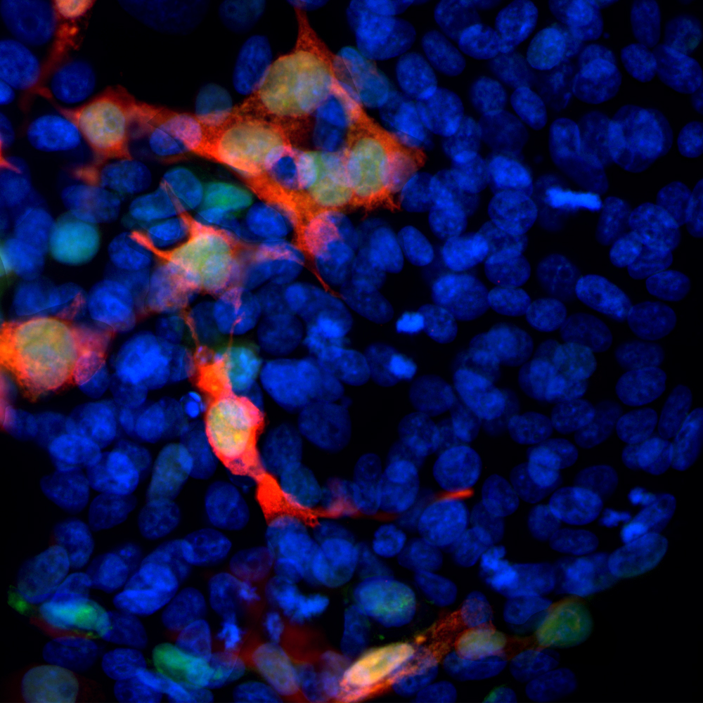
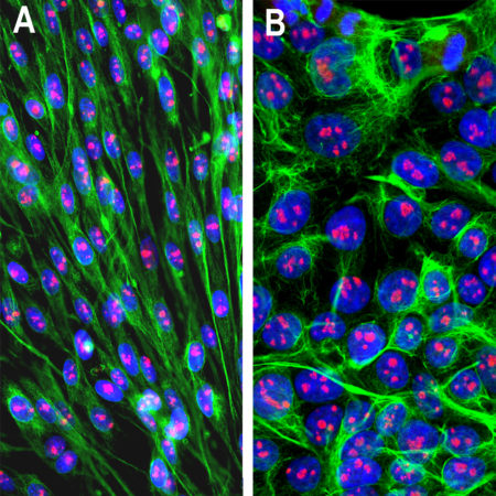
Immunofluorescent analysis of transfected HEK293 cells with a GFP-construct in green stained with mouse mAb to GFP, MCA-3B11, dilution 1:1,000 in red. The blue is Hoechst staining of nuclear DNA. MCA-3B11 antibody reveals GFP protein expressed only in transfected cells, as a result cells are appeared in orange-golden color.
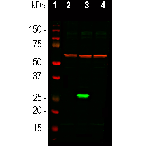
Western blot analysis of HEK293 cell lysates using mouse mAb to GFP, MCA-3B11, in green, dilution 1:1,000: [1] protein standard, [2] non-transfected control cells, [3] cells transfected with a GFP construct, and [4] cells transfected with mCherry construct. The strong green band at ~27kDa corresponds to GFP protein detected only in cells transfected with GFP construct, the antibody does not recognize mCherry. The same blot was simultaneously probed with chicken pAb to HSP60, CPCA-HSP60, dilution 1:10,000, in red. The single band at 60kDa represents HSP60 protein expressed in all preparations.
Mouse Monoclonal Antibody to GFP
Cat# MCA-3B11
$120.00 – $800.00
The green fluorescent protein (GFP) is a 27kDa protein isolated originally from the jellyfish Aequoria victoria. It has an endogenous fluorochrome activity with excitation maximum at 395nm and emission maximum at 509nm, which is similar to that of fluorescein (1,2). The GFP gene was sequenced and the origin of the fluorochrome by autocatalytic activity of certain amino acids was discovered (3,4). Much interest in GFP was generated when it was shown that fluorescence develops rapidly when the protein is expressed and requires only molecular oxygen and no other cofactors. As a result GFP can be expressed in fluorescent form in essentially any prokaryotic or eukaryotic cell (5). GFP has been engineered to produce a vast number of variously colored mutants including blue, cyan and yellow protein derivatives, BFP, CFP and YFP (6-9). GFP and other fluorescent proteins derived from other Cnidarians (jellyfish, coral and medusa) are widely used as tracers in transfection and transgenic experiments to monitor gene expression and protein localization in vivo and in in vitro. The crystal structure of GFP was determined (7) which allowed amino acid modifications to improve spectral properties and prevent multimerization (8,9). The discovery GFP was the basis of the 2008 Nobel prize in chemistry, specifically “for the discovery and development of the green fluorescent protein, GFP”.
The MCA-3B11 antibody was made against a recombinant GFP construct originating from an Aequoria species which was engineered to improve spectral properties and prevent oligomerization (10). This form of GFP, referred to as AcGFP, is 94% identical to the eGFP developed by Tsien and coworkers and is the form of GFP inserted in the Clontech/Takara pAcGFP and related expression vectors. We epitope mapped this antibody to the N-terminal 18 amino acids of the AcGFP protein, the peptide MVSKGAELFTGIVPILIE, which is found in the Takara/Clontech and other GFP vectors and distinct from the sequence seen in other fluorescent proteins. The homologous region of eGFP is MVSKGEELFTGVVPILVE, and this antibody binds this peptide also. We also supply the immunogen, PROT-AcGFP. The antibody can be used to verify the expression, size and stability of both AcGFP and eGFP fusion proteins in western blotting experiments and to amplify GFP signals in tissues of transgenic animals. We also supply another mouse monoclonal antibody with a different isotype and rabbit, chicken, goat polyclonal antibodies to this protein, MCA-3B11, RPCA-GFP, CPCA-GFP and GPCA-GFP.Mouse select image above left for larger view.
AcGFP used as immunogen for this antibody
Sequence taken from AY233272, download here
1 MVSKGAELFT GIVPILIELN GDVNGHKFSV SGEGEGDATY GKLTLKFICT TGKLPVPWPT
61 LVTTLSYGVQ CFSRYPDHMK QHDFFKSAMP EGYIQERTIF FEDDGNYKSR AEVKFEGDTL
121 VNRIELTGTD FKEDGNILGN KMEYNYNAHN VYIMTDKAKN GIKVNFKIRH NIEDGSVQLA
181 DHYQQNTPIG DGPVLLPDNH YLSTQSALSK DPNEKRDHMI YFGFVTAAAI THGMDELYK
This construct is available as PROT-r-AcGFP
1. Shimomura O, Johnson FH, Saiga Y. Extraction, purification and properties of aequorin, a bioluminescent protein from the luminous hydromedusan, Aequorea. J. Cell. Comp. Physiol. 3:223–39 (1962).
2. Shimomura, O. Structure of the chromophore of Aequorea green fluorescent protein. FEBS Lett. 104:220–2 (1979).
3. Prasher DC, et al. Primary structure of the Aequorea victoria green-fluorescent protein. Gene 111:229-33 (1992).
4. Cody CW, et al. Chemical structure of the hexapeptide chromophore of the Aequorea green-fluorescent protein. Biochem. 32:1212-8 (1993).
5. Chalfie M, et al. Green Fluorescent protein as a marker for gene expression. Science 263:802-5 (1994).
6. Heim R, Prasher DC, Tsien RY. Wavelength mutations and post-translational autoxidation of green fluorescent protein. PNAS 91:12501-04 (1994).
7. Ormo M, et al. Crystal structure of the Aequorea victoria green fluorescent protein. Science 273:1392-95 (1996).
8. Tsien RY. The green fluorescent protein. Annu. Rev. Biochem. 67:509-44 (1998).
9. Zacharias DA, Violin JD, Newton AC, Tsien RY. Partitioning of lipid-modified monomeric GFPs into membrane microdomains of live cells. Science 296:913-6 (2002).
10. Gurskaya NG, et al. A colourless green fluorescent protein homologue from the non-fluorescent hydromedusa Aequorea coerulescens and its fluorescent mutants. Biochem. J. 373:403-8 (2003).